

Case Reports
doi: 10.1102/1470-7330.2006.0030.
MRI features of intrahepatic extramedullary haematopoiesis in sickle cell anaemia
Affiliations
- PMID: 17208673
- PMCID: PMC1766560
- DOI: 10.1102/1470-7330.2006.0030
Item in Clipboard
Case Reports
MRI features of intrahepatic extramedullary haematopoiesis in sickle cell anaemia
Cancer Imaging.
.
Abstract
Extramedullary haematopoiesis (EMH) is a reactive mechanism by which blood cells are produced outside of the bone marrow to supplement insufficient production or increased destruction of erythrocytes. EMH is uncommon in sickle cell anaemia (SCA). We report the first case of focal intra-hepatic EMH in SCA depicted on MRI occurring in a 32-year-old woman with homozygote SCA and in view of previously published data, highlight the diagnostic features suggesting a differential diagnosis with other focal liver lesions including infectious, inflammatory or primary liver tumors.
(c) International Cancer Imaging Society.
Figures
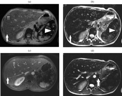
A 32-year-old woman with homozygote sickle cell disease and a 15-year history of transfusion. The liver shows diffuse low signal intensity on all sequences. (a) Fat saturation GRE images show the presence of multiple nodules hyperintense within the liver and remaining isointense to the muscle (arrow and double arrows). Nodules displaying a similar pattern of signal intensity are identified in the spleen (arrowhead). (b) T2 WI TSE fat saturation images show that the multiple nodes are slightly hyperintense to the adjacent liver, and isointense to the muscle (arrow). (c) Following injection of Gd-DOTA (Dotarem ® , Guerbet, Aulnay, France), previously identified nodes show absent enhancement on arterial phase and moderate enhancement on portal phase imaging (arrow and double arrows). Nodules displaying a similar pattern of signal intensity are identified in the spleen (arrowhead). (d) Multiple para-aortic nodes showing low intensity on TSE T2 WI (arrow) consistent with iron accumulation were discovered incidentally.
References
-
- Wong Y, Chen F, Tai KS, et al. Imaging features of focal intrahepatic extramedullary haematopoiesis. Br J Radiol. 1999;72:906–10. - PubMed
-
- Navarro M, Crespo C, Perez L, Martinez C, Galant J, Gonzalez I. Massive intrahepatic extramedullary hematopoiesis in myelofibrosis. Abdom Imaging. 2000;25:184–6. - PubMed
-
- Lonergan GJ, Cline DB, Abbondanzo SL. Sickle cell anemia. Radiographics. 2001;21:971–94. - PubMed
-
- Kushner JP, Porter JP, Olivieri NF. Secondary iron overload. Hematology (Am Soc Hematol Educ Program) 2001:47–61. - PubMed
-
- Kumar A, Aggarwal S, de Tilly LN. Case of the season. Thalassemia major with extramedullary hematopoiesis in the liver. Semin Roentgenol. 1995;30:99–101. - PubMed
Publication types
MeSH terms
LinkOut - more resources
Full Text Sources
Medical