. 2007 Mar;189(6):2487-96.
doi: 10.1128/JB.01491-06. Epub 2007 Jan 5.

A type IV-secretion-like system is required for conjugative DNA transport of broad-host-range plasmid pIP501 in gram-positive bacteria

Affiliations

A type IV-secretion-like system is required for conjugative DNA transport of broad-host-range plasmid pIP501 in gram-positive bacteria

Mohammad Y Abajy et al. J Bacteriol. 2007 Mar.

Abstract

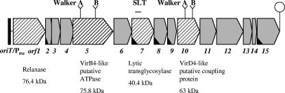
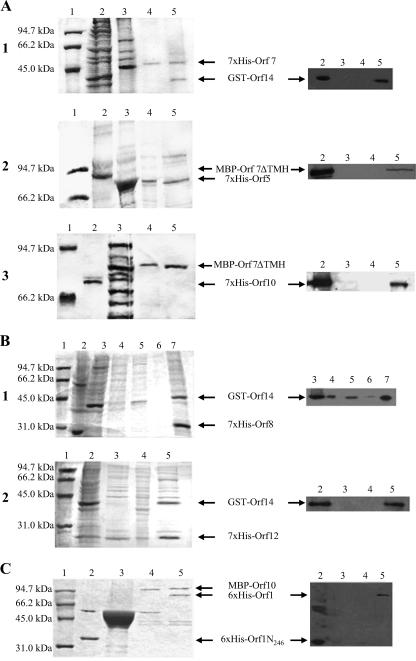
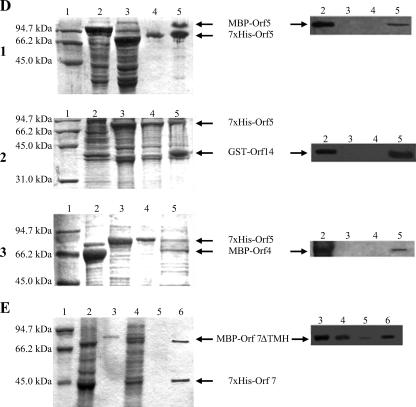
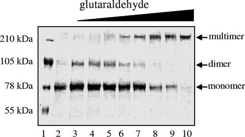
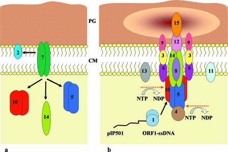
Plasmid pIP501 has a very broad host range for conjugative transfer among a wide variety of gram-positive bacteria and gram-negative Escherichia coli. Functionality of the pIP501 transfer (tra) genes in E. coli was proven by pIP501 retrotransfer to Enterococcus faecalis (B. Kurenbach, C. Bohn, J. Prabhu, M. Abudukerim, U. Szewzyk, and E. Grohmann, Plasmid 50:86-93, 2003). The 15 pIP501 tra genes are organized in a single operon (B. Kurenbach, J. Kopeć, M. Mägdefrau, K. Andreas, W. Keller, C. Bohn, M. Y. Abajy, and E. Grohmann, Microbiology 152:637-645, 2006). The pIP501 tra operon is negatively autoregulated at the transcriptional level by the conjugative DNA relaxase TraA. Three of the 15 pIP501-encoded Tra proteins show significant sequence similarity to the Agrobacterium type IV secretion system proteins VirB1, VirB4, and VirD4. Here we report a comprehensive protein-protein interaction map of all of the pIP501-encoded Tra proteins determined by the yeast two-hybrid assay. Most of the interactions were verified in vitro by isolation of the protein complexes with pull-down assays. In conjunction with known or postulated functions of the pIP501-encoded Tra proteins and computer-assisted prediction of their cellular location, we propose a model for the first type IV-secretion-like system encoded by a conjugative plasmid from gram-positive bacteria.


Figures

FIG. 1.
FIG. 1.
Organization of the pIP501 tra region. The overlapping oriT and Ptra regions are indicated (not to scale). The operon (orf1 to orf15) is terminated by a putative strong rho-independent termination signal (hairpin). Hatched segments indicate genes encoding proteins similar to those identified in G− bacterial T4SSs. The Orf5 protein shows conserved features, a nucleotide binding site motif A (Walker A box 250GLSGGGKT257) and a motif B (Walker B box 509DEFHFLL515), of proteins belonging to the VirB4 family of nucleoside triphosphate-binding proteins (COG3451). Orf10 is a member of the pfam02534 family of TraG/TrwB/TraD/VirD4 coupling proteins. It shows the P-loop motif (Walker A box) and a Walker B motif for nucleotide binding. Orf7 (a VirB1 homolog of the Agrobacterium T-DNA transfer system) contains the SLT domain present in bacterial lytic transglycosylases and was shown to cleave peptidoglycan isolated from E. faecalis and E. coli in an in vitro muramidase assay (Söllü and Grohmann, unpublished data). Open reading frames whose corresponding gene products contain potential signal peptide sequences are marked with a black wedge within the segment.
FIG. 2.
FIG. 2.
In vitro binding assays. (A to E) SDS-PAGE gels (left panels) and corresponding Western blots (right panels). (A) Interactions probably involved in recruitment of Tra proteins to the T4SLS complex. (A1) 12% SDS-PAGE. Lane 1, low-range protein standard (Bio-Rad); lane 2, GST-Orf14 lysate; lane 3, 7×His-Orf7 lysate; lane 4, negative control 7×His-Orf7/GST; lane 5, eluate of the GST-Orf14-7×His-Orf7 complex. Corresponding Western blot with anti-GST antibodies. (A2) 6% SDS-PAGE. Lane 1, low-range protein standard; lane 2, MBP-Orf7ΔTMH lysate; lane 3, 7×His-Orf5 lysate; lane 4, negative control 7×His-Orf5/MBP; lane 5, eluate of the 7×His-Orf5-MBP-Orf7ΔTMH complex. Corresponding Western blot with anti-MBP antibodies. (A3) 8% SDS-PAGE. Lane 1, low-range protein standard; lane 2, 7×His-Orf10; lane 3, MBP-Orf7ΔTMH lysate; lane 4, negative control MBP-Orf7ΔTMH/pQTEV lysate; lane 5, eluate of the MBP-Orf7ΔTMH-7×His-Orf10 complex. Corresponding Western blot with anti-Penta-His antibodies.(B) Interactions of postulated core complex components. (B1) 12% SDS-PAGE. Lane 1, low-range protein standard; lane 2, 7×His-Orf8 lysate; lane 3, GST-Orf14 lysate; lane 4, proteins not bound to Ni-NTA column; lanes 5 and 6, wash steps after binding of the protein complex; lane 7, eluate of the 7×His-Orf8-GST-Orf14 complex. Corresponding Western blot with anti-GST antibodies. (B2) 12% SDS-PAGE. Lane 1, low-range protein standard; lane 2, 7×His-Orf12 lysate; lane 3, GST-Orf14 lysate; lane 4, negative control 7×His-Orf12/GST; lane 5, eluate of the 7×His-Orf12-GST-ORF14 complex. Corresponding Western blot with anti-GST antibodies. (C) Interactions of the putative coupling protein Orf10 in a 12% SDS-PAGE gel. Lane 1, low-range protein standard; lane 2, 6×His-Orf1N246; lane 3, negative control MBP/6×His-Orf1N246; lane 4, eluate of the MBP-Orf10-6×His-Orf1N246 complex; lane 5, eluate of the MBP-Orf10-6×His-Orf1 complex. Corresponding Western blot with anti-Penta-His antibodies. (D) Interactions of the putative ATPase ORF5. (D1) 12% SDS-PAGE. Lane 1, low-range protein standard; lane 2, MBP-Orf5 lysate; lane 3, 7×His-Orf5 lysate; lane 4, negative control 7×His-Orf5/MBP; lane 5, eluate of the 7×His-ORF5-MBP-ORF5 complex. Corresponding Western blot with anti-MBP antibodies. (D2) 12% SDS-PAGE. Lane 1, low-range protein standard; lane 2, GST-Orf14 lysate; lane 3, 7×His-Orf5 lysate; lane 4, negative control 7×His-Orf5/GST; lane 5, eluate of the 7×His-Orf5-GST-Orf14 complex. Corresponding Western blot with anti-GST antibodies. (D3) 10% SDS-PAGE. Lane 1, low-range protein standard; lane 2, MBP-Orf4 lysate; lane 3, 7×His-Orf5 lysate; lane 4, negative control 7×His-Orf5/MBP; lane 5, eluate of the 7×His-Orf5-MBP-Orf4 complex. Corresponding Western blot with anti-MBP antibodies. (E) Homotypic interaction of the lytic transglycosylase Orf7 in a 12% SDS-PAGE. Lane 1, low-range protein standard; lane 2, 7×His-Orf7 lysate; lane 3, purified MBP-Orf7ΔTMH; lane 4, proteins not bound to Ni-NTA column; lane 5, wash step after binding of the protein complex; lane 6, eluate of the 7×His-Orf7-MBP-Orf7ΔTMH complex. Corresponding Western blot with anti-MBP antibodies.
FIG. 2.
FIG. 2.
In vitro binding assays. (A to E) SDS-PAGE gels (left panels) and corresponding Western blots (right panels). (A) Interactions probably involved in recruitment of Tra proteins to the T4SLS complex. (A1) 12% SDS-PAGE. Lane 1, low-range protein standard (Bio-Rad); lane 2, GST-Orf14 lysate; lane 3, 7×His-Orf7 lysate; lane 4, negative control 7×His-Orf7/GST; lane 5, eluate of the GST-Orf14-7×His-Orf7 complex. Corresponding Western blot with anti-GST antibodies. (A2) 6% SDS-PAGE. Lane 1, low-range protein standard; lane 2, MBP-Orf7ΔTMH lysate; lane 3, 7×His-Orf5 lysate; lane 4, negative control 7×His-Orf5/MBP; lane 5, eluate of the 7×His-Orf5-MBP-Orf7ΔTMH complex. Corresponding Western blot with anti-MBP antibodies. (A3) 8% SDS-PAGE. Lane 1, low-range protein standard; lane 2, 7×His-Orf10; lane 3, MBP-Orf7ΔTMH lysate; lane 4, negative control MBP-Orf7ΔTMH/pQTEV lysate; lane 5, eluate of the MBP-Orf7ΔTMH-7×His-Orf10 complex. Corresponding Western blot with anti-Penta-His antibodies.(B) Interactions of postulated core complex components. (B1) 12% SDS-PAGE. Lane 1, low-range protein standard; lane 2, 7×His-Orf8 lysate; lane 3, GST-Orf14 lysate; lane 4, proteins not bound to Ni-NTA column; lanes 5 and 6, wash steps after binding of the protein complex; lane 7, eluate of the 7×His-Orf8-GST-Orf14 complex. Corresponding Western blot with anti-GST antibodies. (B2) 12% SDS-PAGE. Lane 1, low-range protein standard; lane 2, 7×His-Orf12 lysate; lane 3, GST-Orf14 lysate; lane 4, negative control 7×His-Orf12/GST; lane 5, eluate of the 7×His-Orf12-GST-ORF14 complex. Corresponding Western blot with anti-GST antibodies. (C) Interactions of the putative coupling protein Orf10 in a 12% SDS-PAGE gel. Lane 1, low-range protein standard; lane 2, 6×His-Orf1N246; lane 3, negative control MBP/6×His-Orf1N246; lane 4, eluate of the MBP-Orf10-6×His-Orf1N246 complex; lane 5, eluate of the MBP-Orf10-6×His-Orf1 complex. Corresponding Western blot with anti-Penta-His antibodies. (D) Interactions of the putative ATPase ORF5. (D1) 12% SDS-PAGE. Lane 1, low-range protein standard; lane 2, MBP-Orf5 lysate; lane 3, 7×His-Orf5 lysate; lane 4, negative control 7×His-Orf5/MBP; lane 5, eluate of the 7×His-ORF5-MBP-ORF5 complex. Corresponding Western blot with anti-MBP antibodies. (D2) 12% SDS-PAGE. Lane 1, low-range protein standard; lane 2, GST-Orf14 lysate; lane 3, 7×His-Orf5 lysate; lane 4, negative control 7×His-Orf5/GST; lane 5, eluate of the 7×His-Orf5-GST-Orf14 complex. Corresponding Western blot with anti-GST antibodies. (D3) 10% SDS-PAGE. Lane 1, low-range protein standard; lane 2, MBP-Orf4 lysate; lane 3, 7×His-Orf5 lysate; lane 4, negative control 7×His-Orf5/MBP; lane 5, eluate of the 7×His-Orf5-MBP-Orf4 complex. Corresponding Western blot with anti-MBP antibodies. (E) Homotypic interaction of the lytic transglycosylase Orf7 in a 12% SDS-PAGE. Lane 1, low-range protein standard; lane 2, 7×His-Orf7 lysate; lane 3, purified MBP-Orf7ΔTMH; lane 4, proteins not bound to Ni-NTA column; lane 5, wash step after binding of the protein complex; lane 6, eluate of the 7×His-Orf7-MBP-Orf7ΔTMH complex. Corresponding Western blot with anti-MBP antibodies.
FIG. 3.
FIG. 3.
Glutaraldehyde cross-linking of 7×His-Orf10. Samples of 0.5 mg/ml of 7×His-Orf10 were incubated with increasing glutaraldehyde concentrations. The products were loaded onto a 10% SDS polyacrylamide gel, electrophoresed at a constant voltage of 180 V, and stained with Coomassie brilliant blue. Lane 1, SeeBlue plus2-prestained protein standards (Invitrogen, Karlsruhe, Germany); lane 2, no glutaraldehyde; lanes 3 to 10, glutaraldehyde at 0.002% (lane 3). 0.003% (lane 4), 0.004% (lane 5), 0.005% (lane 6), 0.006% (lane 7), 0.007% (lane 8), 0.008% (lane 9); and 0.01% (lane 10).
FIG. 4.
FIG. 4.
Working model for pIP501 conjugative transfer. The postulated DNA secretion complex is assembled in a manner reminiscent of a simplified T4SS. Arrows indicate protein-protein interactions determined with the yeast two-hybrid system. Protein localization is consistent with computer predictions made by using Psort (22), PHDhtm (42), HMMTOP (50, 51), TMPred (28), TMAP (39, 40), and TopPred (16, 52) (available at www.expasy.org). Decreased shading of peptidoglycan (PG) symbolizes Orf7-mediated local opening of PG. (a) Protein-protein interactions detected for Orf7. (b) Assembly of the putative pIP501 transport apparatus. Numbers refer to proteins specified by the pIP501 tra region. Dashed arrows mark putative ATPases. CM, cytoplasmic membrane; ssDNA, single-stranded DNA; NTP, nucleoside triphosphate; NDP, nucleoside diphosphate.

References

    1. Atmakuri, K., E. Cascales, and P. J. Christie. 2004. Energetic components VirD4, VirB11 and VirB4 mediate early DNA transfer reactions required for bacterial type IV secretion. Mol. Microbiol. 54:1199-1211. - PMC - PubMed
    1. Averhoff, B. 2004. DNA transport and natural transformation in mesophilic and thermophilic bacteria. J. Bioenerg. Biomembr. 36:25-33. - PubMed
    1. Bannam, T. L., W. L. Teng, D. Bulach, D. Lyras, and J. I. Rood. 2006. Functional identification of conjugation and replication regions of the tetracycline resistance plasmid pCW3 from Clostridium perfringens. J. Bacteriol. 188:4942-4951. - PMC - PubMed
    1. Baron, C., M. Llosa, S. Zhou, and P. C. Zambryski. 1997. VirB1, a component of the T-complex transfer machinery of Agrobacterium tumefaciens, is processed to a C-terminal secreted product, VirB1*. J. Bacteriol. 179:1203-1210. - PMC - PubMed
    1. Bayer, M., R. Eferl, G. Zellnig, K. Teferle, A. Dijkstra, G. Koraimann, and G. Högenauer. 1995. Gene 19 of plasmid R1 is required for both efficient conjugative DNA transfer and bacteriophage R17 infection. J. Bacteriol. 177:4279-4288. - PMC - PubMed

Publication types

MeSH terms

LinkOut - more resources