. 2007 Jan;19(1):46-62.
doi: 10.1105/tpc.106.047506. Epub 2007 Jan 5.

The indeterminate gametophyte1 gene of maize encodes a LOB domain protein required for embryo Sac and leaf development

Affiliations

The indeterminate gametophyte1 gene of maize encodes a LOB domain protein required for embryo Sac and leaf development

Matthew M S Evans. Plant Cell. 2007 Jan.

Abstract

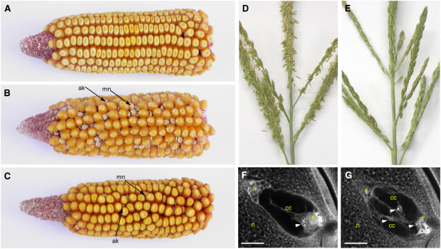
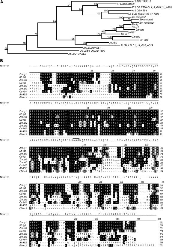
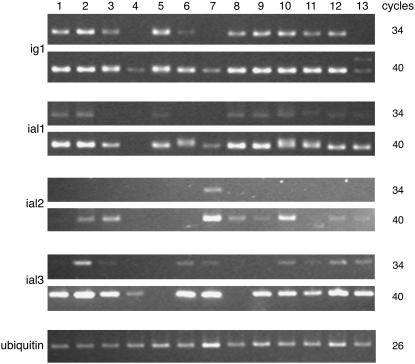
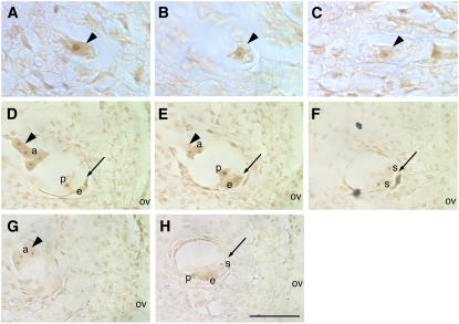
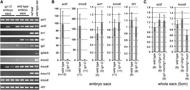
Angiosperm embryo sac development begins with a phase of free nuclear division followed by cellularization and differentiation of cell types. The indeterminate gametophyte1 (ig1) gene of maize (Zea mays) restricts the proliferative phase of female gametophyte development. ig1 mutant female gametophytes have a prolonged phase of free nuclear divisions leading to a variety of embryo sac abnormalities, including extra egg cells, extra polar nuclei, and extra synergids. Positional cloning of ig1 was performed based on the genome sequence of the orthologous region in rice. ig1 encodes a LATERAL ORGAN BOUNDARIES domain protein with high similarity to ASYMMETRIC LEAVES2 of Arabidopsis thaliana. A second mutant allele of ig1 was identified in a noncomplementation screen using active Mutator transposable element lines. Homozygous ig1 mutants have abnormal leaf morphology as well as abnormal embryo sac development. Affected leaves have disrupted abaxial-adaxial polarity and fail to repress the expression of meristem-specific knotted-like homeobox (knox) genes in leaf primordia, causing a proliferative, stem cell identity to persist in these cells. Despite the superficial similarity of ig1-O leaves and embryo sacs, ectopic knox gene expression cannot be detected in ig1-O embryo sacs.


Figures

Figure 1.
Figure 1.
Phenotypes of ig1 Mutants. (A) Ear from a W64A wild-type plant. (B) Ear from an ig1-O/ig1-O W64A mutant plant. (C) Ear from an ig1-mum/ig1-mum W64A plant. (D) Fertile wild-type tassel with anthers extruded. (E) Sterile ig1/ig1 mutant tassel without extruded anthers. (F) Wild-type embryo sac. (G) ig1 embryo sac. Asterisks indicate degenerated synergids. Arrowheads point to polar nuclei. a, antipodal cells; ak, aborted kernel; cc, central cell; e, egg cell; mn, miniature kernel; n, nucellus. Bars = 25 μm.
Figure 2.
Figure 2.
Comparative Mapping between Rice and Maize around ig1. BAC clones and maize BAC contigs are stippled. Arrows indicate the physical distance between markers on rice chromosome 1. Vertical lines show the positions of the closest sequence matches in the rice genome for maize clones in the ig1 region. Maize markers shown above the maize BACs had been placed on the BAC clones but not on the genetic map. Underlines indicate PCR-based markers designed using rice genome information.
Figure 3.
Figure 3.
ig1 Gene Structure and Mutations. (A) Structures of the ig1 gene and its rice ortholog. Positions of the Mu insertion in ig1-mum and the Hopscotch insertion in ig1-O are indicated. The LOB domain is indicated by dark gray. The first exon contains most of the 5′ untranslated region (UTR); the second exon contains the LOB domain; the third exon contains the C-terminal domain; and the fourth exon contains most of the 3′ untranslated region. The third intron is 1.6 kb in length and extends beyond the end of AZM4_49905. The last exon is part of a separate AZM contig, AZM4_116957. (B) Effect of ig1 mutations on ig1 RNA levels. RT-PCR was performed on RNA from whole ear primordia ∼5 cm in length using primers for both ig1 around the first intron and for ubiquitin (ubi). Lane 1, wild type; lane 2, ig1-mum homozygote; lane 3, ig1-O homozygote. Homozygous mutants were identified on the basis of male sterility. (C) Relative ig1 message levels in ig1-O and ig1-mum ear primordia normalized to ubi. The left graph shows PCR results using primers around the first intron of ig1 (around the insertion in ig1-mum and upstream of the insertion in ig1-O), and the right graph shows PCR results using primers around the second intron of ig1 (downstream of the insertion in ig1-mum and around the insertion in ig1-O). Error bars indicate se. Five replicate assays were performed for each pair of PCR primers per sample.
Figure 4.
Figure 4.
The ig1 Gene Family. (A) Relationship of IG1 with other closely related LOB domain proteins. Only the portion of the LOB domain protein family including AS2 and its five most closely related homologs from Arabidopsis is shown. Of the rice and maize genes and the full-length LOB domain genes from a few other species, only those that fall into this same LOB domain subfamily are included. The number above each branch corresponds to the posterior probability for that node. (B) Alignment of proteins within the IG1/AS2 subgroup. Residues identical to IG1 are highlighted. The LOB domain is indicated by a black line over the residues, and the SKY motif is boxed. The translation start of the P. taeda gene is unknown. Zm, Zea mays; Sb, Sorghum bicolor; Os, Oryza sativa; At, Arabidopsis thaliana; Pt, Pinus taeda; Cs, Citrus sinensis.
Figure 5.
Figure 5.
Expression of ig1 and ial Genes in Various Tissues. RT-PCR was performed on total RNA using ubi primers or ig1 or ial primers. Note the larger ig1 transcript seen after 40 cycles in ig1-mum mutant leaves (lane 13). Lane 1, fully expanded leaf; lane 2, immature leaf; lane 3, husk leaf; lane 4, root; lane 5, silk; lane 6, endosperm at 9 d after pollination; lane 7, mature pollen; lane 8, 1-cm-long ear primordium; lane 9, 5-cm-long ear primordium; lane 10, 2-cm-long tassel primordium; lane 11, ovule; lane 12, ligular region of wild-type seedling leaf; lane 13, ligular region of ig1-mum flag leaf.
Figure 6.
Figure 6.
Expression of ig1 in Floral Organs. (A) Diagram of a maize female spikelet with a fully developed upper floret and an arrested lower floret (yellow). In a median section, the lemma and palea (the first-whorl organs), one of the arrested stamens, and the silk (gynoecium) with a single ovule can be seen. The area in box 1 is shown in (B), (C), and (D). The area in box 2 is shown in (E). The area in box 3 is shown in (F) and (G). The area in box 4 is shown in Figures 7A to 7C. The area in box 5 is shown in Figures 7D to 7H. (B) and (D) Median longitudinal sections of wild-type ovules. (C) Longitudinal section through the margin of a wild-type ovule. ig1 is expressed in the adaxial epidermis at the base of the carpels that make up the silk as well as at the boundary between the integuments and the nucellus. (E) The lower floret and the palea of the upper floret. (F) and (G) The lemma of the upper floret and the inner glume (bract). Probes were as follows: (B), (C), (E), and (F), ig1 antisense probe; (D) and (G), ig1 sense probe. Arrowheads point to adaxial expression of ig1 in floral organs and bracts. Arrows point to embryo sacs. g, glume; i, integument; l, lemma; n, nucellus; p, palea; si, silk; st, stamen. Bar = 100 μm.
Figure 7.
Figure 7.
Expression of ig1 in Embryo Sacs. (A) to (C) Ovules with stage 1 (one-nucleus) embryo sacs. (A) and (C) Wild-type stage 1 embryo sacs (arrowheads). The ig1 antisense probe detects message in stage 1 embryo sacs. (B) Nonfunctional megaspore (arrowhead) lacking ig1 expression from the same ovule as the embryo sac in (A). (D) to (H) Ovules with immature cellularized embryo sacs. Arrowheads point to chalazal nuclei and cells of embryo sacs, and arrows point to micropylar nuclei and cells of embryo sacs. Probes were as follows: (A), (B), (D), (E), and (F), ig1 antisense probe; (C), (G), and (H), ig1 sense probe. Signal with the ig1 antisense probe is higher than that with the sense probe control in the antipodal cells. In the cells at the micropylar end of the embryo sac, the signal is more variable and the background staining with the sense probe is higher, making it difficult to determine whether there is any ig1 signal. a, antipodal cell; e, egg; ov, ovary wall; p, polar nucleus; s, synergid. Bar = 25 μm for (A) to (C) and 50 μm for (D) to (H).
Figure 8.
Figure 8.
Phenotype of the Flag Leaf (Last Vegetative Leaf) in ig1/ig1 Homozygotes. (A) and (C) Wild type. (B), (D), (F), and (G) ig1-mum/ig1-mum. (E) ig1-O/ig1-O W23. (A) and (B) Adaxial side of the middle of the leaf blade. (C) to (E) Adaxial ligular region. The arrow points to the leaf flap along the midrib. Arrowheads point to the ligular region. The leaf in (B) shows the most common phenotype in ig1-mum plants. The leaf in (D) shows the most severe ig1-mum leaf phenotype. The leaf in (E) shows the most common ig1-O flag leaf phenotype in a W23 inbred background, with leaf flaps on the sheath and mild ligule distortion. (F) Close-up of leaf flaps on the adaxial surface of an ig1-mum leaf. Note that there are epidermal hairs on the outer epidermis of the flaps but not the inner surfaces of the flaps. The edges of the flaps also have hairs, like normal leaf margins. (G) Knot protruding from the abaxial surface of an ig1-mum flag leaf, with anthocyanin and abaxial epidermal hairs typical of sheath tissue. (H) Expression of knox genes in ig1-mum and rs2. PCR was performed on cDNA from the ligular region of a wild-type flag leaf (lane 1), the ligular region of an ig1-mum flag leaf (lane 2), the ligular region of wild-type seedling leaves (lane 3), the ligular region of rs2 seedling leaves (lane 4), ear primordia (lane 5), or genomic DNA (lane 6).
Figure 9.
Figure 9.
Polarity Defects in ig1 Flag Leaves. (A) Midrib of a wild-type flag leaf. The arrowhead points to abaxial sclerenchyma. (B) Midrib and flaps of an ig1-mum flag leaf. Arrowheads point to abaxial and adaxial sclerenchyma. (C) Normal leaf margin. Arrowhead points to marginal sclerenchyma. (D) Margin of an ig1-mum leaf flap. Arrowhead points to marginal sclerenchyma. (E) Model of adaxial and abaxial domains in wild-type and ig1 leaves. cc, clear cells; ph, phloem; x, xylem. Bars = 150 μm.
Figure 10.
Figure 10.
Expression of Genes from Whole Embryo Sacs. RNA was subjected to linear amplification and then used for quantitative real-time PCR. (A) Products after the completion of real-time RT-PCR. (B) Comparison of RNA levels of act2, knox8, act1, knox6, and rld1 in wild-type and ig1-O embryo sacs. Expression of ubi was used to normalize RNA levels between samples. Expression is given relative to that of ig1-O. Because of the dramatic difference between ig1-O and wild-type embryo sacs for act2 and knox8, these genes are compared using a log scale, whereas all others are presented on a linear scale. (C) Relative levels of RNA of act2 and knox8 in developing wild-type, ig1-O, and ig1-mum ear primordia. Error bars in (B) and (C) represent se. The number of replicates is given in parentheses below each column.

References

    1. Acharya, A., Ruvinov, S.B., Gal, J., Moll, J.R., and Vinson, C. (2002). A heterodimerizing leucine zipper coiled coil system for examining the specificity of a position interactions: amino acids I, V, L, N, A, and K. Biochemistry 41 14122–14131. - PubMed
    1. Banks, J.A. (1999). Gametophyte development in ferns. Annu. Rev. Plant Physiol. Plant Mol. Biol. 50 163–186. - PubMed
    1. Bortiri, E., Chuck, G., Vollbrecht, E., Rocheford, T., Martienssen, R., and Hake, S. (2006). ramosa2 encodes a LATERAL ORGAN BOUNDARY domain protein that determines the fate of stem cells in branch meristems of maize. Plant Cell 18 574–585. - PMC - PubMed
    1. Byrne, M.E., Barley, R., Curtis, M., Arroyo, J.M., Dunham, M., Hudson, A., and Martienssen, R.A. (2000). Asymmetric leaves1 mediates leaf patterning and stem cell function in Arabidopsis. Nature 408 967–971. - PubMed
    1. Cove, D.J., and Knight, C.D. (1993). The moss Physcomitrella patens, a model system with potential for the study of plant reproduction. Plant Cell 5 1483–1488. - PMC - PubMed

Publication types

MeSH terms

Associated data