. 2007 Jan 16;46(2):483-91.
doi: 10.1021/bi061935g.

Redox-induced changes in flavin structure and roles of flavin N(5) and the ribityl 2'-OH group in regulating PutA--membrane binding

Affiliations

Redox-induced changes in flavin structure and roles of flavin N(5) and the ribityl 2'-OH group in regulating PutA--membrane binding

Weimin Zhang et al. Biochemistry. .

Abstract

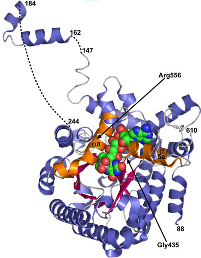
PutA is a novel flavoprotein in Escherichia coli that switches from a transcriptional repressor to a membrane-bound proline catabolic enzyme. Previous crystallographic studies of the PutA proline dehydrogenase (PRODH) domain under oxidizing conditions revealed that FAD N(5) and the ribityl 2'-OH group form hydrogen bonds with Arg431 and Arg556, respectively. Here we identify molecular interactions in the PutA PRODH active site that underlie redox-dependent functional switching of PutA. We report that reduction of the PRODH domain induces major structural changes in the FAD cofactor, including a 22 degrees bend of the isoalloxazine ring along the N(5)-N(10) axis, crankshaft rotation of the upper part of the ribityl chain, and formation of a new hydrogen bond network involving the ribityl 2'-OH group, FAD N(1), and Gly435. The roles of the FAD 2'-OH group and the FAD N(5)-Arg431 hydrogen bond pair in regulating redox-dependent PutA-membrane associations were tested using FAD analogues and site-directed mutagenesis. Kinetic membrane binding measurements and cell-based reporter gene assays of modified PutA proteins show that disrupting the FAD N(5)-Arg431 interaction impairs the reductive activation of PutA-membrane binding. We also show that the FAD 2'-OH group acts as a redox-sensitive toggle switch that controls PutA-membrane binding. These results illustrate a new versatility of the ribityl chain in flavoprotein mechanisms.


Figures

FIGURE 1
FIGURE 1
Ribbon drawing of dithionite-reduced PutA86-669. β-strands of the β8α8 barrel are colored pink and α-helices are colored blue. The two helices α8 and α5a are highlighted in orange. The FAD cofactor (green) and bound SO22- (gray/red) are drawn in CPK mode. The dashed curves represent disordered residues. Selected residue numbers are indicated.
FIGURE 2
FIGURE 2
Three views of the active site of dithionite-reduced PutA86-669. A) Highlighting interactions with SO22-, B) conformation of FAD 2′-OH, and C) nonplanar isoalloxazine. In each panel, the red dashed lines denote electrostatic interactions (hydrogen bonds, ion pairs) and the blue cage represents a simulated annealing σA-weighted mFo-DFc electron density map contoured at 3.5σ.
FIGURE 3
FIGURE 3
Stereo view of conformational differences between dithionite-reduced PutA86-669 and oxidized ligand-bound PutA86-669. Reduced PutA86-669 with bound SO22- is shown in white. Oxidized PutA86-669 with bound substrate analogue L-THFA is colored green. Black dotted lines indicate hydrogen bonds observed in both structures. Green dotted lines indicate the hydrogen bond between Arg556 and 2′-OH observed only in the oxidized enzyme. Orange dotted lines indicate hydrogen bonds unique to the reduced enzyme.
FIGURE 4
FIGURE 4
SPR sensorgrams of reduced PutA proteins injected onto a L1 chip coated with E. coli polar lipid vesicles. Sensorgrams (a) 100 nM reconstituted FAD PutA (5 mM dithionite), (b) 100 nM R431M (5 mM dithionite), and (c) 100 nM 5-deaza-FAD PutA (50 mM dithionite), respectively. PutA proteins were injected at 60 μL/min in the presence of sodium dithionite in HEPES-N buffer (pH 7.4) with arrows indicating the beginning and end of the protein sample injection. No lipid interactions are observed with R431M and 5-deaza-FAD PutA.
FIGURE 5
FIGURE 5
Proline-dependent transcriptional activation of the putC:lacZ reporter gene. β-galactosidase activity from E. coli strain JT31 putA lacZ containing the putC:lacZ reporter construct and either pUC18 empty vector (■), pUC18-PutA (●), or pUC18-PutA R431M (♦) grown in medium supplemented with increasing amounts of proline. Also shown is β-galactosidase activity from cells containing putC:lacZ and pUC18-PutA grown in medium supplemented with increasing amounts of LTHFA (▲). Bars represent the ± standard errors of the mean from at least three independent experiments. Solid curve is the fit of the increase in β-galactosidase activity as a function of proline concentration in cells containing pUC18-PutA to the equation Pobs = Pmax * [pro]/(Keq + [pro]).
FIGURE 6
FIGURE 6
SPR kinetic analyses of oxidized 2′-deoxy-FAD-PutA, oxidized PutA R556M and reduced PutA R556M binding to E. coli polar lipids at 25 °C in HEPES-N buffer (pH 7.4). Sensorgrams of increasing concentrations of A) oxidized 2′-deoxy-FAD-PutA (12.5, 25, 50, 100, and 200 nM) and B) oxidized PutA R556M (6.25, 12.5, 25, 50, and 100 nM). C) From bottom to top, sensorgrams of increasing concentrations of PutA R556M (6.25, 12, 25, 50, and 100 nM) in the presence of 5 mM sodium dithionite. For all sensorgrams, association phase (1) corresponds to injection of the PutA protein at 60 μl/min for 120 s and the dissociation phase (2) corresponds to the flow of HEPES-N buffer at 60 μl/min for 300 s. The data were fit by global analysis to 1:1 Langmuir binding isotherm. Signals from the control surface have been subtracted.

Similar articles

Cited by

References

    1. Brown E, Wood JM. Redesigned purification yields a fully functional PutA protein dimer from Escherichia coli. J. Biol. Chem. 1992;267:13086–13092. - PubMed
    1. Menzel R, Roth J. Enzymatic properties of the purified putA protein from Salmonella typhimurium. J. Biol. Chem. 1981;256:9762–9766. - PubMed
    1. Menzel R, Roth J. Regulation of genes for proline utilization in Salmonella typhimurium: autogenous repression by the putA gene product. J. Mol. Biol. 1981;148:21–44. - PubMed
    1. Vílchez S, Manzanera M, Ramos J. Control of expression of divergent Pseudomonas putida put promoters for proline catabolism. Appl. Environ. Microbiol. 2000;66:5221–5225. - PMC - PubMed
    1. Lee YH, Nadaraia S, Gu D, Becker DF, Tanner JJ. Structure of the proline dehydrogenase domain of the multifunctional PutA flavoprotein. Nat. Struct. Biol. 2003;10:109–114. - PMC - PubMed

Publication types

MeSH terms

Associated data

LinkOut - more resources