Review
. 2007 Feb 1;401(3):623-33.
doi: 10.1042/BJ20061062.

Structural and functional analysis of the Na+/H+ exchanger

Affiliations
Review

Structural and functional analysis of the Na+/H+ exchanger

Emily R Slepkov et al. Biochem J. .

Abstract

The mammalian NHE (Na+/H+ exchanger) is a ubiquitously expressed integral membrane protein that regulates intracellular pH by removing a proton in exchange for an extracellular sodium ion. Of the nine known isoforms of the mammalian NHEs, the first isoform discovered (NHE1) is the most thoroughly characterized. NHE1 is involved in numerous physiological processes in mammals, including regulation of intracellular pH, cell-volume control, cytoskeletal organization, heart disease and cancer. NHE comprises two domains: an N-terminal membrane domain that functions to transport ions, and a C-terminal cytoplasmic regulatory domain that regulates the activity and mediates cytoskeletal interactions. Although the exact mechanism of transport by NHE1 remains elusive, recent studies have identified amino acid residues that are important for NHE function. In addition, progress has been made regarding the elucidation of the structure of NHEs. Specifically, the structure of a single TM (transmembrane) segment from NHE1 has been solved, and the high-resolution structure of the bacterial Na+/H+ antiporter NhaA has recently been elucidated. In this review we discuss what is known about both functional and structural aspects of NHE1. We relate the known structural data for NHE1 to the NhaA structure, where TM IV of NHE1 shows surprising structural similarity with TM IV of NhaA, despite little primary sequence similarity. Further experiments that will be required to fully understand the mechanism of transport and regulation of the NHE1 protein are discussed.


Figures
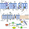
Figure 1
Figure 1. Model of NHE1 showing the membrane and cytoplasmic domains
Upper panel: topology of the membrane domain which functions to transport cations. This illustration is based on the findings of Wakabayashi et al. [90]. Pink circles, residues implicated in both ion transport and inhibitor binding; orange circles, residues implicated in ion binding and transport; yellow circles, residues implicated in inhibitor binding, green circles, residues implicated in NHE1 folding and targeting to the plasma membrane. Lower panel: representation of the cytoplasmic domain which functions to regulate the membrane domain through interactions with signalling molecules.
Figure 2
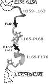
Figure 2. Structure of a TM IV peptide in a membrane -mimetic environment
Convergent stretches (grey) of residues Asp159–Leu163, Leu165–Pro168, and Ile169–Phe176 in relation to pivot residues Phe176 and Pro168/Ile169 are shown [137]. The flexible N- and C-termini are represented by dashed lines. Note that only the proline side chains are indicated. Reproduced from [4] with permission.
Figure 3
Figure 3. Structure of the E. coli Na+/H+ antiporter NhaA
Ribbon representation of NhaA viewed in parallel with the membrane. The 12 TM segments are labelled with Roman numerals. TM segments IV and XI have a helix-extended chain–helix conformation. The cytoplasmic (upper) and periplasmic (lower) faces of the membrane are indicated by broken lines. TM segments are colour coded as follows: I, pink; II, cyan; III, blue; IV, red; V, grey; VI, green; VII, yellow; VIII, orange; IX, green; X, pale yellow; XI, brown; XII cyan. This Figure is adapted from [112] with permission from Nature © 2005 Macmillan Magazines Ltd. (http://www.nature.com/).
Figure 4
Figure 4. Proposed mechanism of transport by the E. coli Na+/H+ antiporter NhaA
The TM IV–TM XI assembly and its interaction with TM IX is shown. (A) Acidic pH-locked conformation. TM IX is bent, and the conformation of the TM IV–TM XI assembly only partly exposes the Na+-binding site. (B) Alkaline pH causes a conformational change in helix IX that results in a reorientation of the TM IV–TM XI assembly. This exposes the Na+-binding site (yellow circle) to the cytoplasmic funnel (red broken lines and red circle) and blocks it from the periplasm (orange line). (C) Na+ binding causes the cation-loaded binding site to be exposed to the periplasm. Upon release of the cation, key aspartic acid residues are protonated, shifting NhaA back into the cytoplasm-exposed conformation in (B). This Figure is adapted from [112] with permission from Nature © 2005 Macmillan Magazines Ltd. (http://www.nature.com/).
Figure 5
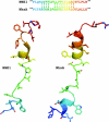
Figure 5. Structural similarity between TM IV segments of NHE1 and NhaA
Upper panel: sequence alignment of TM IVs of NHE1 and NhaA with arbitrary coloration. Lower panel: representative NHE1 TM IV structure [4] (arbitrary orientation at swivel point Phe164) shown alongside the TM IV segment of NhaA [112], with colouring as indicated in the sequence alignment. Despite little sequence similarity, alignment of the TM IV segments of NHE1 and NhaA over the residues illustrated allows structural superimposition at the 14 pairs of residues indicated by arrows. Differences in structure between the Leu163–Phe164 swivel point of NHE1 and the crystal structure of inactive NhaA at Ile128–Pro129 mean that the entire segment does not superimpose well. However, Asp159–Phe162 of NHE1 shows extremely similar structure to Ile121–Trp126 of NhaA, and a subset of the NMR structures with the appropriate Pro168/Ile169 swivel point orientation gives excellent superimposition of Leu165–Gly174 of NHE1 on Ale130–Gly139 of NhaA.

References

    1. Orlowski J., Grinstein S. Na+/H+ exchangers of mammalian cells. J. Biol. Chem. 1997;272:22373–22376. - PubMed
    1. Karmazyn M., Gan T., Humphreys R. A., Yoshida H., Kusumoto K. The myocardial Na+–H+ exchange. Structure, regulation, and its role in heart disease. Circ. Res. 1999;85:777–786. - PubMed
    1. Cardone R. A., Casavola V., Reshkin S. J. The role of disturbed pH dynamics and the Na+/H+ exchanger in metastasis. Nat. Rev. Cancer. 2005;5:786–795. - PubMed
    1. Slepkov E. R., Rainey J. K., Li X., Liu Y., Cheng F. J., Lindhout D. A., Sykes B. D., Fliegel L. Structural and functional characterization of transmembrane segment IV of the NHE1 isoform of the Na+/H+ exchanger. J. Biol. Chem. 2005;280:17863–17872. - PubMed
    1. Orlowski J., Grinstein S. Diversity of the mammalian sodium/proton exchanger SLC9 gene family. Pflügers Arch. 2004;447:549–565. - PubMed

Publication types

MeSH terms

Substances