

Case Reports
doi: 10.1186/1750-1172-2-2.
Craniocervical junction malformation in a child with Oromandibular-limb hypogenesis-Möbius syndrome
Affiliations
- PMID: 17210070
- PMCID: PMC1774563
- DOI: 10.1186/1750-1172-2-2
Item in Clipboard
Case Reports
Craniocervical junction malformation in a child with Oromandibular-limb hypogenesis-Möbius syndrome
Orphanet J Rare Dis.
.
Abstract
We report a male child with Oromandibular-limb hypogenesis (OMLH), the main features being bilateral sixth and seventh nerve palsies, limb anomalies and hypoplasia of the tongue. Additional features were shortness of the neck associated with torticollis. Radiographs of the cervical spine were non-contributory, but 3D computed tomography (CT) scanning of this area identified: a) congenital hypoplasia of the atlas; b) the simultaneous development of occiput-atlas malformation/developmental defect. To our knowledge, this is the first clinical report assessing the cervico-cranium malformation in a child with OMLH-Möbius syndrome.
Figures
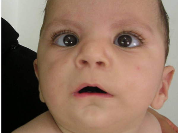
Proband phenotype.
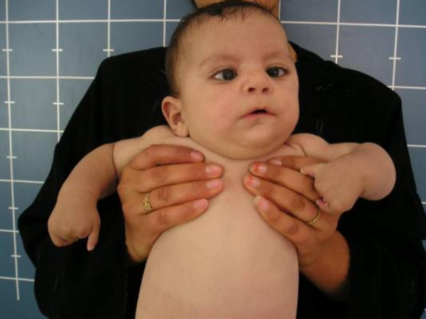
Proband phenotype and hands: Subtotal absence of the phalanges (preservation of the hypoplastic thumbs and hypoplastic 5th fingers, respectively).
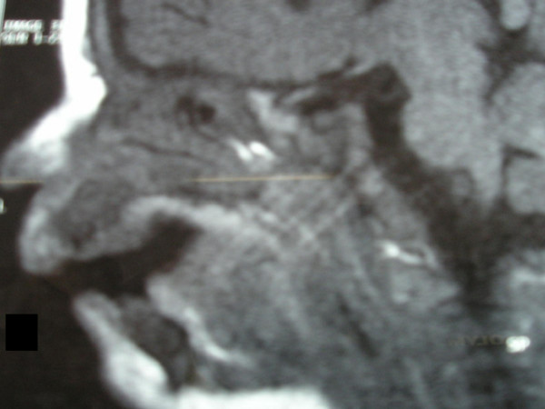
Sagittal MRI imaging, showed markedly hypoplastic tongue.
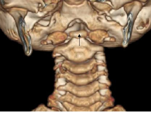
3D reconstruction CT scan ; Hypoplastic anterior arch of the atlas and the impacted os terminale of the odontoid (arrow) between the two halves of the maldeveloped anterior arch of the atlas-the os terminale usually fuses at 12 years of age-this can be confused with fracture.
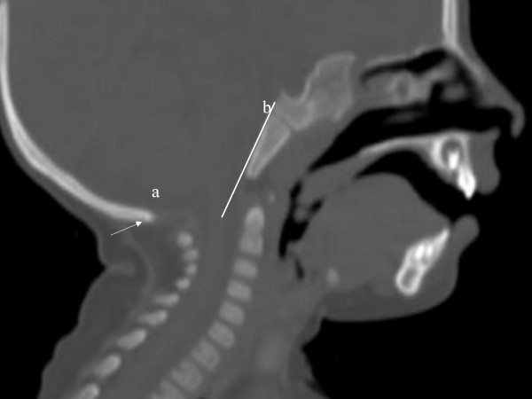
3D sagittal CT scan; Agenesis of the posterior arch of the atlas (arrow-a). Arrow (b) notes the Wachenheim clivus line, which is drawn along the posterior aspect of the clivus toward the odontoid process; in our patient the line does not intersect or is it tangential to the odontoid process. The latter confirms the existence of progressive craniocervical abnormality.
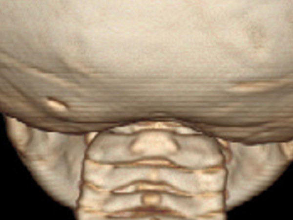
3D reconstruction CT scan showed agenesis of posterior arch of the atlas.
Similar articles
-
Oromandibular limb hypogenesis syndrome with no oromandibular features, or Moebius syndrome without facial palsy? A diagnostic conundrum.Clin Dysmorphol. 2009 Apr;18(2):107-109. doi: 10.1097/MCD.0b013e3283244396. Clin Dysmorphol. 2009. PMID: 19305193 No abstract available.
-
Oromandibular limb hypogenesis complex (Hanhart syndrome): a severe adult phenotype.Am J Med Genet. 1999 Apr 23;83(5):427-9. Am J Med Genet. 1999. PMID: 10232758 No abstract available.
-
Craniofacial phenotypes associated with Robinow syndrome.Am J Med Genet A. 2021 Dec;185(12):3606-3612. doi: 10.1002/ajmg.a.61986. Epub 2020 Nov 25. Am J Med Genet A. 2021. PMID: 33237614
-
[Oromandibular-limb hypogenesis spectrum].Ryoikibetsu Shokogun Shirizu. 2000;(30 Pt 5):224-5. Ryoikibetsu Shokogun Shirizu. 2000. PMID: 11057206 Review. Japanese. No abstract available.
-
[Partial trisomy 10q syndrome].Ryoikibetsu Shokogun Shirizu. 2000;(30 Pt 5):325-6. Ryoikibetsu Shokogun Shirizu. 2000. PMID: 11057247 Review. Japanese. No abstract available.
Cited by
-
Occipito-vertebral dissociation in connection with extensive cervical spine malsegmentation in a boy with Möbius syndrome.Clinics (Sao Paulo). 2009;64(10):1034-6. doi: 10.1590/S1807-59322009001000016. Clinics (Sao Paulo). 2009. PMID: 19841713 Free PMC article. No abstract available.
-
A novel malformation complex of bilateral and symmetric preaxial radial ray-thumb aplasia and lower limb defects with minimal facial dysmorphic features: a case report and literature review.Cases J. 2008 Oct 24;1(1):271. doi: 10.1186/1757-1626-1-271. Cases J. 2008. PMID: 18950501 Free PMC article.
References
-
- Harris JH, Mirvis SE. The radiology of acute cervical spine trauma. 3. Baltimore, Md: Williams & Wilkins; 1996. The normal cervical spine; pp. 1–73.
-
- Hall BD. Aglossia-adactylia. Birth Defects Orig Artic Ser. 1971;7:233–236. - PubMed
-
- Hanhart E. Ueber die Kombination von Peromelie mit Mikrognathie, ein neues Syndrom beim Menschen, entsprechend der Akroteriasis congenita von Wriedt und Mohr beim Rind. Arch Klaus-Stift Ver. 1950;25:531–543.
Publication types
MeSH terms
LinkOut - more resources
Full Text Sources
Medical
