. 2007 May;19(5):1034-43.
doi: 10.1016/j.cellsig.2006.11.010. Epub 2006 Nov 28.

Activated Ras induces cytoplasmic vacuolation and non-apoptotic death in glioblastoma cells via novel effector pathways

Affiliations

Activated Ras induces cytoplasmic vacuolation and non-apoptotic death in glioblastoma cells via novel effector pathways

Aparna Kaul et al. Cell Signal. 2007 May.

Abstract

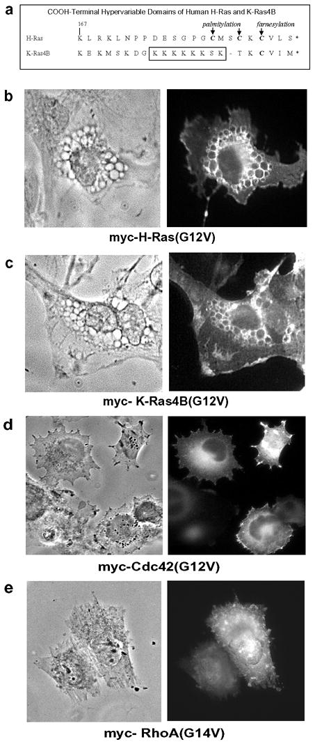
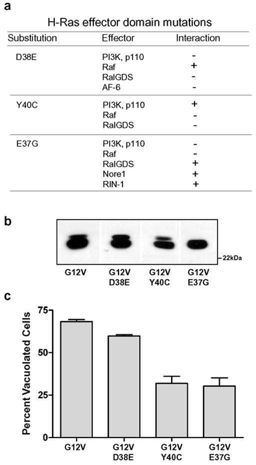
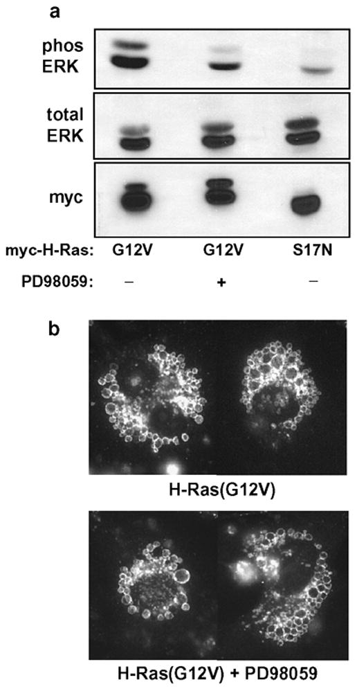
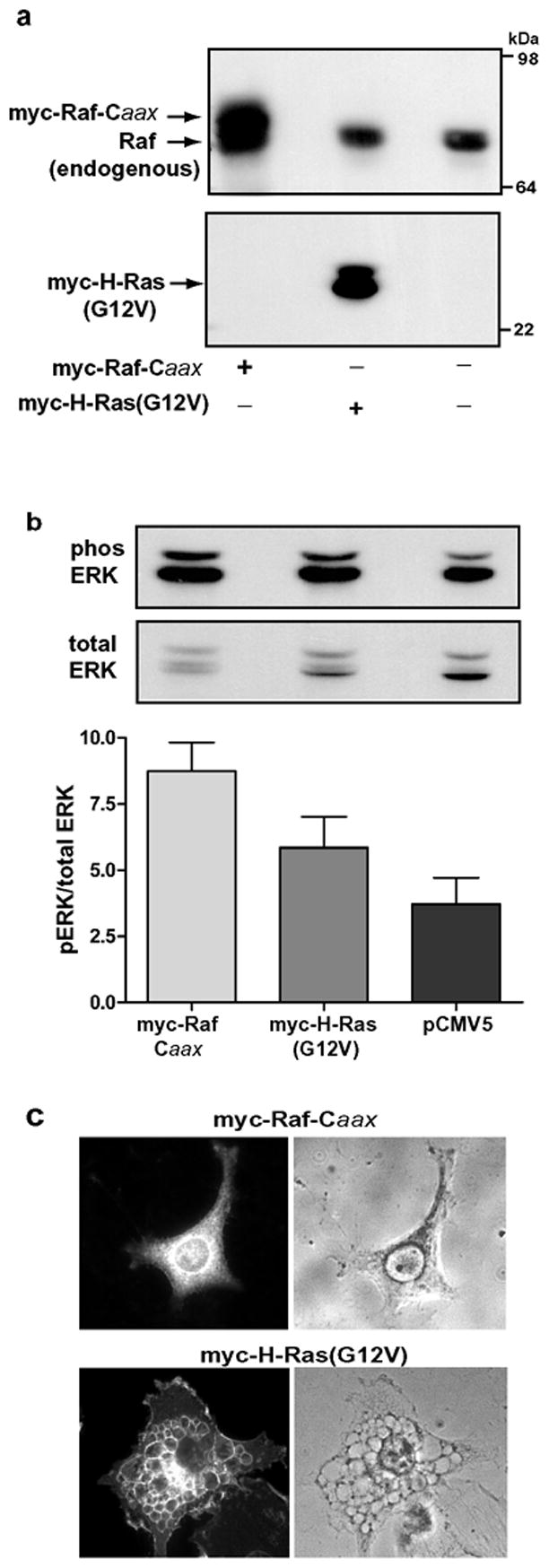
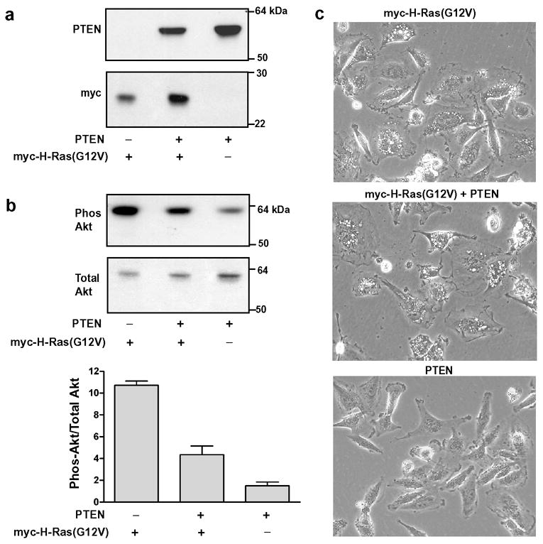
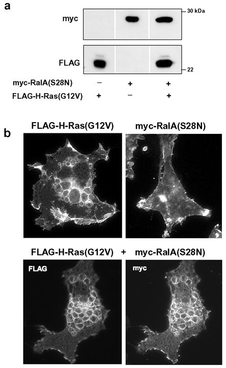
Expression of activated H-Ras induces a unique form of non-apoptotic cell death in human glioblastoma cells and other specific tumor cell lines. The major cytopathological features of this form of death are the accumulation of large phase-lucent, LAMP1-positive, cytoplasmic vacuoles. In this study we sought to determine if induction of cytoplasmic vacuolation a) depends on Ras farnesylation, b) is specific to H-Ras, and c) is mediated by signaling through the major known Ras effector pathways. We find that the unusual effects of activated H-Ras depend on farnesylation and membrane association of the GTPase. Both H-Ras(G12V) and K-Ras4B(G12V) stimulate vacuolation, but activated forms of Cdc42 and RhoA do not. Amino acid substitutions in the Ras effector domain, which are known to selectively impair its interactions with Raf kinase, class-I phosphatidylinositide 3-kinase (PI3K), or Ral nucleotide exchange factors, initially pointed to Raf as a possible mediator of cell vacuolation. However, the MEK inhibitor, PD98059, did not block the induction of vacuoles, and constitutively active Raf-Caax did not mimic the effects of Ras(G12V). Introduction of normal PTEN together with H-Ras(G12V) into U251 glioblastoma cells reduced the PI3K-dependent activation of Akt, but had no effect on vacuolation. Finally, co-expression of H-Ras(G12V) with a dominant-negative form of RalA did not suppress vacuolation. Taken together, the observations indicate that Ras activates non-conventional and perhaps unique effector pathways to induce cytoplasmic vacuolation in glioblastoma cells. Identification of the relevant signaling pathways may uncover specific molecular targets that can be manipulated to activate non-apoptotic cell death in this type of cancer.


Figures

Figure 1
Figure 1
Stimulation of vacuole formation by activated Ras depends on farnesylation. (a) U251 glioblastoma cells were nucleofected with expression vectors encoding the indicated myc-tagged constructs. After 24 hours equal amounts of cellular protein were subjected to western blot analysis using the myc antibody. At the same time, live cells in parallel cultures were examined by phase contrast microscopy (300 × magnification). (b) U251 cells were nucleofected with expression vectors encoding the indicated myc-tagged constructs. After 24 hours the expressed proteins were localized by immunofluorescence microscopy as described in Materials and methods. The left panel of each figure shows the same cells under phase contrast.
Figure 2
Figure 2. H- and K-Ras, but not Rho family GTPases, induce vacuolation.
(a) Sequence alignment of the COOH-terminal hypervariable domains of H-Ras and K-Ras4B, showing sites of posttranslational lipidation. (b–e) U251 glioblastoma cells were nucleofected with expression vectors encoding the indicated myc-tagged constructs. After 24 hours the expressed proteins were localized by immunofluorescence microscopy using an antibody against the myc epitope (right panel). The left panel of each figure shows the same field of cells under phase contrast.
Figure 3
Figure 3
Mutations in the effector domain of H-Ras(G12V) have differential effects on its ability to induce cytoplasmic vacuolation. (a) Summary of Ras effector interactions known to be inhibited (−) or permitted (+) by specific amino acid substitutions, based on information compiled from refs ,–,,,. (b) U251 cells were nucleofected with expression vectors encoding the indicated myc-tagged H-Ras constructs. After 24h, equal aliquots of cell protein were subjected to western blot analysis using the myc antibody (representative blot shown). (c) Parallel cultures were processed for immunofluorescence localization of myc-tagged Ras proteins. Cells staining positive with the myc antibody were photographed and scored for vacuolation. Cells were scored as vacuolated if they contained at least 2 vacuoles ≥ 0.5 μ in diameter and/or >10 smaller vacuoles. At least 65 myc-positive cells were scored to determine the percentage of transfected cells that were vacuolated in each culture. The results for each Ras construct shown in the bar graph are derived from three separate experiments (mean ± S.D.). The decreases in vacuolation observed with G12V+Y40C and G12V+E37G were significant at p<0.01 compared with G12V alone (Student’s t-test).
Figure 4
Figure 4
Induction of vacuoles by H-Ras(G12V) does not depend on activation of the ERK pathway. To obtain uniform expression of exogenous Ras, U251 cells were infected with retrovirus encoding myc-H-Ras(G12V) or myc-H-Ras(S17N). At the time that the cells were plated in selection medium (see Methods), the MEK inhibitor, PD98059 (25 μM) or an equivalent volume of DMSO vehicle, was added to the culture medium. Cells were processed for western blot analysis or immunofluorescent detection of LAMP1-positive vacuoles after 42 h in the presence of the inhibitor. (a) Activation of ERK was monitored in equal aliquots of whole cell lysate (normalized for protein) using antibodies to detect phosphorylated ERK1/2 (phos ERK) and total ERK1/2 . (b) Immunofluorescence microscopy to detect LAMP1-positive vacuoles was performed as described in Materials and methods. The panels are composites showing representative cells from the cultures incubated with or without PD98059.
Figure 5
Figure 5
Overexpression of constitutively active Raf-1 does not mimic the effect of activated H-Ras. U251 cells were nucleofected with empty vector or with expression vectors encoding myc-Raf-Caax or myc-H-Ras(G12V). The cells were processed for western blot analysis or immunofluorescence 24 h later. (a) Immunoblot analysis with an antibody against Raf confirms that myc-Raf-Caax (upper band) is overexpressed relative to endogenous Raf. (b) Phospho-ERK and total ERK were determined by immunoblot analysis (representative blot shown). The ratio of phospho-ERK to total ERK was determined by quantifying the immunoblot signals with a Kodak Image station. The results shown in the bar graph are the means (±SD) of separate determinations performed on three cultures. (c) Cells expressing myc-Raf-Caax or myc-H- Ras(G12V) were identified by immunofluorescence using antibody against the myc epitope. The same cells were examined under phase contrast to assess the presence of vacuoles. The nucleofection efficiencies in both sets of cultures were approximately 50%. There were no vacuolated myc-positive cells in the cultures expressing myc-Raf-Caax.
Figure 6
Figure 6
Reduction of the activity of the PI 3K signaling pathway by introduction of PTEN does not prevent the induction of vacuoles by H-Ras(G12V). (a) U251 cells were nucleofected with expression vectors encoding PTEN and/or myc-H-Ras(G12V) as indicated. After 24 h equal aliquots of cell protein were processed for western blot analysis to verify the expression of PTEN and myc-tagged Ras. (b) Phospho-Akt and total Akt were determined by immunoblot analysis (representative blot shown). The bar graph depicts the ratio of phospho-Akt to total Akt (mean ± S.D.) determined by quantifying the immunoblot signals from three separate cultures. The decrease in Akt phosphorylation in PTEN + myc-H-Ras(G12V) was significant at p < 0.05 compared with myc-H-Ras(G12V) alone (Student’s t-test). (c) Phase contrast images of representative fields of cells from the cultures harvested for the blots shown in a & b (200x).
Figure 7
Figure 7
Dominant-negative RalA does not block the induction of vacuolation by activated H-Ras. (a) U251 cells were nucleofected with expression vectors encoding FLAG-H-Ras(G12V), myc-RalA(S28N) or a combination of both. Equal amounts of cell lysate were subjected to western blot analysis to verify expression of the tagged proteins. (b) The upper panels show representative immunofluorescent localization of FLAG-H-Ras(G12V) alone and myc-RalA(S28N) alone. The bottom panels show a typical cell expressing both proteins.

Similar articles

Cited by

References

    1. Bos JL. ras oncogenes in human cancer: a review. Cancer Res. 1989;49:4682–4689. - PubMed
    1. Campbell SL, Khosravi-Far R, Rossman KL, Clark GJ, Der CJ. Increasing complexity of Ras signaling. Oncogene. 1998;17:1395–1413. - PubMed
    1. Shields JM, Pruitt K, McFall A, Shaub A, Der CJ. Understanding Ras: 'it ain't over 'til it's over'. Trends Cell Biol. 2000;10:147–154. - PubMed
    1. Der CJ, Finkel T, Cooper GM. Biological and biochemical properties of human rasH genes mutated at codon 61. Cell. 1986;44:167–176. - PubMed
    1. Lowy DR, Willumsen BM. Function and regulation of Ras. Annu Rev Biochem. 1993;62:851–891. - PubMed

Publication types

MeSH terms