. 2007 Feb 1;42(3):385-93.
doi: 10.1016/j.freeradbiomed.2006.11.009. Epub 2006 Nov 10.

Accumulation of oxidatively generated DNA damage in the brain: a mechanism of neurotoxicity

Affiliations

Accumulation of oxidatively generated DNA damage in the brain: a mechanism of neurotoxicity

Liuji Chen et al. Free Radic Biol Med. .

Abstract

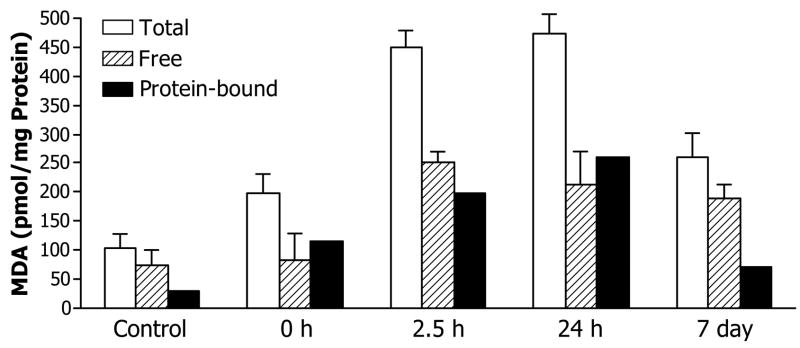
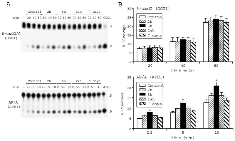
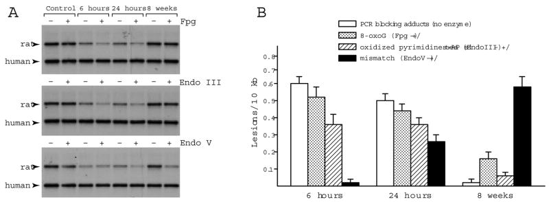
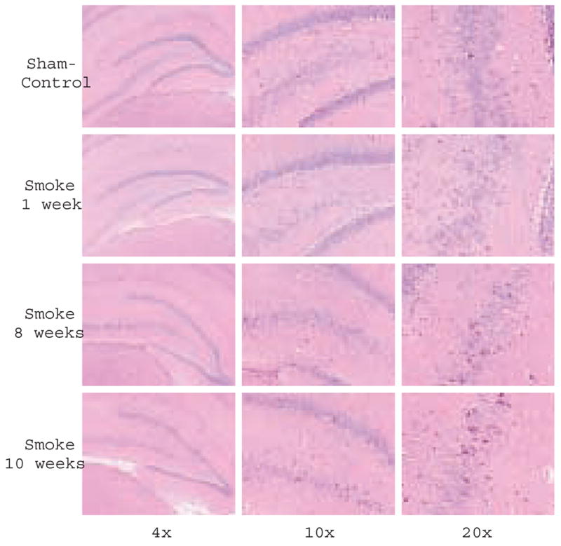
Unrepaired or erroneously repaired DNA lesions drive genomic instability and contribute to cellular and organ decline. Since delayed neuropathologies are common in survivors of smoke inhalation injuries, we asked whether the integrity of brain DNA might be compromised by acute exposure to combustion smoke. Although many studies demonstrate that the brain is equipped to repair oxidatively damaged DNA, to date, the capacity for accurate DNA repair under conditions of disrupted oxygenation and oxidative stress has not been defined. We show that DNA adducts detectable by their ability to block PCR amplification form in the rat hippocampus after acute exposure to smoke. To identify the different types of adducts and to dissect their temporal formation and repair profiles in vivo in the brain, we used DNA-modifying enzymes to convert specific adducts into strand breaks prior to PCR amplification. Using this strategy, we detected formation of oxidative DNA adducts early on after smoke inhalation, while mismatched bases emerged at the later recovery times, potentially due to an erroneous DNA repair process. Erroneous repair can be mutagenic and because the initial smoke-induced oxidative damage to DNA is extensive, compromised fidelity of DNA repair may underlie neurotoxicity and contribute to delayed death of hippocampal neurons.


Figures

Fig 1
Fig 1
Smoke inhalation-induced MDA formation in the brain. Free and total MDA levels were determined; values for protein bound MDA were obtained by subtraction. Values represent pmoles of MDA per mg protein extract expressed as means±SD, n=3.
Fig 2
Fig 2
Smoke inhalation induces 8-oxodG immunoreactivity in the hippocampus. Representative photomicrographs of sections (Bregma -3.14) from control rats and rats harvested at 6, 24 hours and 7 days post smoke stained with the 8-oxodG antibody. Staining is brown and nuclei are counterstained light blue with hematoxylin. Significantly elevated 8-oxodG immunoreactivity is observed at 6 and 24 hours post smoke with a decline in staining by 7 days.
Fig 3
Fig 3
Smoke inhalation modulates BER in the hippocampus. In vitro cleavage of oxidative adducts by hippocampal nuclear extracts from control rats and rats at 2, 6, 24 hours and 7 days post smoke is shown: (A) Autoradiograms of incision products (P) generated over time by cleavage of end labeled double stranded oligonucleotide substrates (S) carrying either the *8-oxodG/C adduct or the abasic site *AP[THF]/A. [−] is a negative control in absence of extract; OGG1 and APE1 lanes show reference reactions assembled with the respective recombinant enzymes. (B) Values from Phosphorimager quantitation of products generated with extracts from 4 rats per group and 3 cleavage assays per extract were averaged and plotted as mean ± SD. * indicates cleavage significantly different from control for each respective time point P<0.05.
Fig 4
Fig 4
Smoke inhalation-induced DNA damage in the rat hippocampus. Representative autoradiograms show PCR products generated by amplification of the MYH gene locus in hippocampal DNA from control rats and rats after exposure to combustion smoke. DNA was preincubated with/without the DNA modifying enzymes, Fpg, Endo III or Endo V, prior to amplification. Amplification levels were reduced at 6 and 24 hours and restored within 8 weeks after exposure to smoke for DNA preincubated without DNA modifying enzymes, reflecting formation and subsequent clearance of the directly arising PCR-blocking adducts. Amplification product of the human β-globin locus (2.2 kb) that serves as an internal control for each PCR reaction is indicated. Amplification levels of DNA preincubated with/without Fpg (+/−) were similar for controls but reduced for rats harvested at 6 and 24 hours post smoke. Likewise, in the case of Endo III, the +/− yields were reduced at 6 and 24 hours and restored by 8 weeks post smoke indicating that levels of Fpg and Endo III targets return to near normal. In contrast, while no reduced amplification was detected for Endo V treated DNA at 6 hours, reduction was seen at 24 hours with a further decrease by 8 weeks, indicative of an increase in the number of Endo V targets. (B) Bar graphs show relative frequencies of adducts generated in hippocampal DNA following smoke inhalation: Relative amplification values for enzyme treated/non-treated DNA were averaged (n=5, triplicate PCR reactions), converted into lesions frequencies and normalized to 10 kb (expressed as mean ± SD). Frequency of directly arising PCR blocking adducts was determined from values obtained following preincubation in absence of DNA modifying enzymes (blank bars). Frequency of sites targeted by Fpg is depicted as dotted bars, by Endo III as hatched bars and Endo V as solid bars.
Fig 5
Fig 5
Delayed neuronal injury after inhalation of smoke. Histochemical analysis of coronal sections from smoke and sham-smoke treated rats: Representative photomicrographs of hematoxylin and eosin staining through the hippocampus (Bregma -3.14) captured with 4x, 10x and 20x objectives, from control rats and rats sacrificed 1, 8 and 10 weeks post smoke exposure. An increase in number of dark neurons (dark pink) is observed at 8 and 10 weeks.

References

    1. Fitzpatrick JC, Cioffie WG., Jr . Diagnosis and treatment of inhalation injury. In: Herndon DN, editor. Total Burn Care, volume. Philadelphia: W.B. Saunders; 1996. pp. 184–192.
    1. Rossi J, 3rd, Ritchie GD, Macys DA, Still KR. An overview of the development, validation, and application of neurobehavioral and neuromolecular toxicity assessment batteries: potential applications to combustion toxicology. Toxicology. 1996;115:107–117. - PubMed
    1. Raub JA, Benignus VA. Carbon monoxide and the nervous system. Neurosci Biobehav Rev. 2002;26:925–940. - PubMed
    1. Weaver LK, Hopkins RO, Chan KJ, Churchill S, Elliott CG, Clemmer TP, Orme JF, Jr, Thomas FO, Morris AH. Hyperbaric oxygen for acute carbon monoxide poisoning. N Engl J Med. 2002;347:1057–1067. - PubMed
    1. Prockop LD. Carbon monoxide brain toxicity: clinical, magnetic resonance imaging, magnetic resonance spectroscopy, and neuropsychological effects in 9 people. J Neuroimaging. 2005;15:144–149. - PubMed

Publication types