

Case Reports
doi: 10.1136/jnnp.2006.109561.
Epub 2007 Jan 8.
A rare symptomatic presentation of ecchordosis physaliphora: neuroradiological and surgical management
Affiliations
- PMID: 17210621
- PMCID: PMC2077974
- DOI: 10.1136/jnnp.2006.109561
Item in Clipboard
Case Reports
A rare symptomatic presentation of ecchordosis physaliphora: neuroradiological and surgical management
J Neurol Neurosurg Psychiatry.
2007 Jun.
Abstract
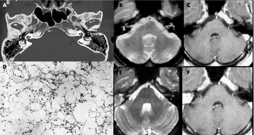
We report a case of ecchordosis physaliphora, an uncommon benign lesion originating from embryonic notochordal remnants, intradurally located in the prepontine cistern, that unusually presented associated with symptoms. MRI detected and precisely located the small mass. At surgery, a cystic gelatinous nodule was found ventral to the pons, contiguous with the dorsal wall of the clivus via a small pedicle. Histological examination diagnosed the lesion as an ecchordosis physaliphora. Here we focus on the analysis of the neuroradiological aspects that play a crucial role from both a diagnostic and a therapeutic standpoint.
Conflict of interest statement
Competing interests: None.
References
-
- Lantos P L, Louis D N, Rosenblum M K.et al Tumours of the nervous system. In: Graham DI, Lantos PL, eds. Greenfield's Neuropathology, vol 2, 7th Edn. London: Arnold, 2002767–1052.
-
- Rodriguez L, Colina J, Lopez J.et al Intradural prepontine growth: giant ecchordosis physaliphora or extraosseous chordoma? Neuropathology 199919336–340.
-
- Toda H, Kondo A, Iwaski K. Neuroradiological characteristics of ecchordosis physaliphora: case report and review of the literature. J Neurosurg 199889830–834. - PubMed
Publication types
MeSH terms
LinkOut - more resources
Full Text Sources