. 2007 Mar;27(6):2324-42.
doi: 10.1128/MCB.02300-06. Epub 2007 Jan 8.

Distinct structural features of caprin-1 mediate its interaction with G3BP-1 and its induction of phosphorylation of eukaryotic translation initiation factor 2alpha, entry to cytoplasmic stress granules, and selective interaction with a subset of mRNAs

Affiliations

Distinct structural features of caprin-1 mediate its interaction with G3BP-1 and its induction of phosphorylation of eukaryotic translation initiation factor 2alpha, entry to cytoplasmic stress granules, and selective interaction with a subset of mRNAs

Samuel Solomon et al. Mol Cell Biol. 2007 Mar.

Abstract

Caprin-1 is a ubiquitously expressed, well-conserved cytoplasmic phosphoprotein that is needed for normal progression through the G(1)-S phase of the cell cycle and occurs in postsynaptic granules in dendrites of neurons. We demonstrate that Caprin-1 colocalizes with RasGAP SH3 domain binding protein-1 (G3BP-1) in cytoplasmic RNA granules associated with microtubules and concentrated in the leading and trailing edge of migrating cells. Caprin-1 exhibits a highly conserved motif, F(M/I/L)Q(D/E)Sx(I/L)D that binds to the NTF-2-like domain of G3BP-1. The carboxy-terminal region of Caprin-1 selectively bound mRNA for c-Myc or cyclin D2, this binding being diminished by mutation of the three RGG motifs and abolished by deletion of the RGG-rich region. Overexpression of Caprin-1 induced phosphorylation of eukaryotic translation initiation factor 2alpha (eIF-2alpha) through a mechanism that depended on its ability to bind mRNA, resulting in global inhibition of protein synthesis. However, cells lacking Caprin-1 exhibited no changes in global rates of protein synthesis, suggesting that physiologically, the effects of Caprin-1 on translation were limited to restricted subsets of mRNAs. Overexpression of Caprin-1 induced the formation of cytoplasmic stress granules (SG). Its ability to bind RNA was required to induce SG formation but not necessarily its ability to enter SG. The ability of Caprin-1 or G3BP-1 to induce SG formation or enter them did not depend on their association with each other. The Caprin-1/G3BP-1 complex is likely to regulate the transport and translation of mRNAs of proteins involved with synaptic plasticity in neurons and cellular proliferation and migration in multiple cell types.


Figures

FIG. 1.
FIG. 1.
Caprin-1 and G3BP-1 associate and colocalize in RNA-rich cytoplasmic granules. (A, B) G3BP-1 coprecipitates with Caprin-1. In panel A, 293T cells were transfected with vector alone or Flag-Caprin-1, and total lysates were subjected to anti-Flag IP. The eluate of the immunoprecipitate (IP) and a 1/10 aliquot of the supernatant remaining after IP (S/N) together with a 1/10 aliquot of the whole-cell lysate (WCL) were run in parallel on SDS-PAGE and blotted for endogenous G3BP-1 with a mouse MAb to G3BP-1. Note that the efficient precipitation of the Flag-Caprin-1 was accompanied by a clearing of G3BP-1 from the post-IP supernatant. Panel B shows a reciprocal experiment in which coprecipitation of endogenous Caprin-1 was detected with anti-Caprin-1 rabbit serum. (C) Colocalization of Caprin-1 and G3BP-1 in cytoplasmic RNA granules. Actively proliferating HeLa cells were stained for endogenous Caprin-1 (green) and G3BP-1 (red). Nuclei were stained with DAPI (blue). The arrows in the enlarged area shown in the inset show colocalization of Caprin-1 and G3BP-1 in cytoplasmic granules. (D) Colocalization of Caprin-1 and RNA in cytoplasmic granules. NIH 3T3 cells were fixed and stained for total cellular RNA using 1 μM ethidium bromide (red) according to the method of Tang et al. (44) and with rabbit anti-Caprin-1 serum (green). As a control, some slides were pretreated with RNase before staining with ethidium bromide and anti-Caprin-1 serum. Note the lack of ethidium bromide staining in the cytoplasmic granules and the nucleoli in the nucleus after RNase treatment.
FIG. 2.
FIG. 2.
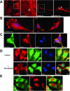
Association of Caprin-1-containing granules with microtubules and with cellular processes. (A) Caprin-1-containing granules occur on a filamentous network resembling the microtubular network and are enriched in cellular processes. 3T3 cells that stably expressed low amounts of Caprin-1-HA were stained for HA (red). Arrows point to the Caprin-1-positive granules arrayed on a filamentous network and enriched in cellular processes. (B, C) Caprin-1 localizes at sites of adhesion and at the leading and trailing edges of migrating 3T3 fibroblasts and HeLa cells. In panel B, 3T3 cells that stably expressed low amounts of Caprin-1-HA were stained for HA (red). In panel C, HeLa cells were stained for endogenous Caprin-1 (green) and G3BP-1 (red). Nuclei were stained with DAPI (blue). The asterisks indicate concentrations of Caprin-1-HA in panel B and Caprin-1 and G3BP-1 in panel C at the sites of cell adhesion and at the leading and trailing edges of cells. (D) Caprin-1-containing cytoplasmic granules are associated with microtubules. Actively growing HeLa cells were treated with nocodazole (33 μM) for 90 min to disrupt microtubules and stained with rabbit anti-Caprin-1 serum (red) and anti-β-tubulin MAb (green). Note that nocodazole treatment resulted in the loss of the filamentous distribution of Caprin-1 in the cytoplasm and the coredistribution of Caprin-1 and β-tubulin into blebs. (E) Caprin-1-containing cytoplasmic granules are transport RNPs. Actively growing HeLa cells were stained with anti-Caprin-1 serum (green) and the stress granule marker TIA-1 (red). Note the absence of TIA-1 in Caprin-1-positive cytoplasmic granules.
FIG. 3.
FIG. 3.
Caprin-1 interacts with G3BP-1 through an evolutionarily conserved peptide motif. (A) G3BP1 binds to a conserved peptide motif in Caprin-1. 293T cells were cotransfected with plasmids expressing Flag-G3BP-1 and various fragments of GFP-Caprin-1 comprising amino acids 1 to 327, 1 to 606, 352 to 606, or 352 to 380, as indicated. Cell lysates were subjected to anti-Flag IP, and precipitated proteins were eluted from the beads and subjected to SDS-PAGE and immunoblotting with anti-GFP, anti-Flag, or anti-G3BP-1 antibodies. (B) (i) Conserved features of Caprin-1- and insect HR-1-containing proteins. The amino-terminal MPSA motifs, the central conserved motif, and the RGG motifs are highlighted. (ii) Also shown is an alignment of the central conserved G3BP-1 binding motif in three insect HR-1-containing proteins and vertebrate Caprin-1 and Caprin-2 together with the core consensus. (C) Peptides containing the G3BP-1-binding Caprin-1 motif compete with Caprin-1 for binding to G3BP-1. (i) Sequence of the core consensus peptide and an extended consensus peptide from the G3BP-1 binding motif from human Caprin-1. (ii) 293T cells were transfected with plasmids expressing Flag-G3BP-1 and GFP-Caprin-1 and lysates were subjected to anti-Flag IP. Washed beads, with bound GFP-Caprin-1 and Flag-G3BP-1 were agitated in 1 ml buffer containing the indicated peptides at 50 μM or 200 μM or buffer alone for 90 min at 4°C. The proteins on the beads were eluted and immunoblotted with anti-GFP and anti-Flag antibodies. (iii) 293T cells were transfected with plasmids expressing Flag-G3BP-1 and GFP-Caprin-1, and lysates were subjected to anti-Flag IP. Washed beads, with bound GFP-Caprin-1 and Flag-G3BP-1 were agitated in 1 ml buffer containing the indicated peptide at 380 μM or buffer alone for 90 min at 4°C. Also shown is a coprecipitation of GFP-Caprin and Flag-G3BP-1, incubated with 100 μg of RNase A in 1 ml of buffer for 60 min. The proteins on the beads were eluted and subjected to SDS-PAGE and immunoblotting with anti-GFP and anti-Flag antibodies.
FIG. 4.
FIG. 4.
G3BP-1 binds Caprin-1 through the NTF-2-like domain. (A) Conservation of the NTF-2-like domain and RNA-binding domain of G3BP-1 in human, Xenopus, and Drosophila cells. (B) G3BP-1 mutants used. (C) The RNA-binding domain of G3BP-1 is not necessary for its interaction with Caprin-1. 293T cells were transfected with plasmids expressing Caprin-1-HA and Flag-G3BP-1 or fragment of G3BP-1 (1 to 340), and the cell lysates were subjected to anti-Flag IP. The proteins on the beads were eluted and subjected to SDS-PAGE and immunoblotting with anti-GFP and anti-Flag antibodies. (D) G3BP-1 binds Caprin-1 through its amino terminus. Cell lysates from 293T cells expressing Flag-Caprin-1 were mixed with bacterially expressed GST-G3BP fragments (1 to 309 and 229 to 466) and subjected to anti-Flag IP. The proteins on the beads were eluted and subjected to SDS-PAGE and immunoblotting with anti-GFP and anti-Flag antibodies. (E) G3BP-1 recognizes Caprin-1 through its NTF-2-like domain. 293T cells were transfected with plasmids expressing Flag-Caprin-1 or Flag-Caprin-1 (47 to 380) and GFP-G3BP-1 (1 to 141), and the cell lysates were subjected to anti-Flag IP. The precipitates were subjected to SDS-PAGE and immunoblotting with anti-GFP and anti-Flag antibody.
FIG. 5.
FIG. 5.
Caprin-1 enters cytoplasmic stress granules and its overexpression induces them. (A, B) Caprin-1 is recruited into SG induced with arsenite. HeLa cells (A) or 3T3 cells that stably expressed low amounts of Caprin-1 HA (B) were stressed by treatment with arsenite (0.5 mM for 1 h). The cells, before or after stress, were fixed and costained for endogenous Caprin-1 (red) and G3BP-1 (green) or with Caprin-1 (green) and TIA-1 (red) for panel A and for HA (red) for panel B. Nuclei were stained with DAPI (blue). Note the recruitment of Caprin-1 and G3BP-1 into cytoplasmic granules which were also positive for TIA-1. Arrows indicate SG in the cytoplasm. (C) Overexpressed Caprin-1 induces cytoplasmic SG containing G3BP-1 and the SG marker TIA-1. HeLa cells transfected with plasmid expressing GFP-Caprin-1 at 48 h were fixed and stained for G3BP-1 (red) or TIA-1 (red). Nuclei were stained with DAPI (blue). (D) Sensitivity of Caprin-1 induced granules to dissolution by treatment with cycloheximide. HeLa cells were transiently transfected with plasmids expressing GFP-Caprin-1 or with GFP-G3BP-1 and at 48 h, and aliquots were treated with 100 μg/ml cycloheximide for 1 h. Note in both cases the disappearance of SG upon treatment with cycloheximide.
FIG. 6.
FIG. 6.
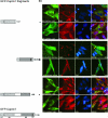
The carboxy-terminal RNA-binding domain of Caprin-1 is necessary for its ability to induce SG when overexpressed. HeLa cells were transfected with plasmid expressing GFP-fusion fragments of Caprin-1, GFP-Caprin-1 (1 to 327), GFP-Caprin-1 (352 to 709), or GFP-Caprin-1 (1 to 606). After 48 h, the cells were fixed and stained for G3BP-1 (red) or TIA-1 (red). Nuclei were stained with DAPI (blue). Note the absence of SG formation by GFP-Caprin-1 (1 to 327) and GFP-Caprin-1 (1 to 606).
FIG. 7.
FIG. 7.
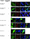
The RNA-binding domain of a single interacting partner of the Caprin-1-G3BP-1 complex is necessary and sufficient for the entry of the complex to SG. (A) HeLa cells were cotransfected with plasmids encoding Flag-G3BP-1 and those expressing GFP fusions of fragments of Caprin-1, namely GFP-Caprin-1 (1 to 606), GFP-Caprin-1 (352 to 606), or GFP-Caprin-1 (1 to 327). After 48 h, the cells were stained for Flag (red). Nuclei were stained with DAPI. None of these Caprin-1 fragments induce SG formation when expressed alone (data not shown). However, when coexpressed with Flag-G3BP-1, the Caprin-1 fragments 1 to 606 and 352 to 606 colocalized with G3BP-1-induced SG, but the Caprin-1 fragment, 1 to 327, that lacks the motif for binding G3BP-1 did not. (B) 293T cells were transfected with plasmid expressing GFP fused to a 29-amino-acid peptide from Caprin-1, GFP-Caprin-1 (352 to 380) with or without Flag-G3BP-1. After 48 h, the cells were fixed and stained for Flag (red). Nuclei were stained with DAPI. Note that the GFP-Caprin-1 (352 to 380) entered SG formed in cells coexpressing G3BP-1. (C) The NTF-2-like domain of G3BP-1 enters SG only when coexpressed with Caprin-1. HeLa cells were transfected with the GFP fusion of the fragment of G3BP-1 corresponding to the NTF-2 like domain, GFP-G3BP-1 (1 to 141), with and without Flag-Caprin-1. After 48 h, the cells were fixed and stained for Flag (red). Nuclei were stained with DAPI. Note that in cells overexpressing Flag-Caprin-1, the GFP-G3BP-1 (1 to 141) relocated from the nucleus into the cytoplasmic SG that were induced by overexpression of Flag-Caprin-1.
FIG. 8.
FIG. 8.
Caprin-1 and G3BP-1 can independently induce the formation of SG and enter them. (A) Caprin-1 is not needed for the formation of SG by G3BP-1 or for the entry of G3BP-1 to SG. Avian R-Caprin-1−/− DT40 cells that express no endogenous Caprin-1 but expressed human Caprin-1 under the control of a DOX-suppressible promoter were grown for 3 days with (+) or without (−) DOX, which suppresses the expression of human Caprin-1 to undetectable levels. They were transfected with GFP-G3BP-1 and grown in the continued presence or absence of 0.5 μg/ml of DOX for 16 h. Note that expression of the GFP-G3BP-1 induced SG (arrows) in both the control cells and those that lacked Caprin-1. (B) Interaction with endogenous G3BP-1 is not needed for Caprin-1 for the formation of SG or for its entry to SG. HeLa cells were transfected with a Flag-tagged fragment of Caprin-1 (381 to 709) that does not interact with G3BP-1, and after 48 h, the cells were fixed and stained. Note the induction of SG containing Flag-Caprin-1 (381 to 709) in the cytoplasm.
FIG. 9.
FIG. 9.
Overexpression of Caprin-1 induces of phosphorylation of eIF-2α through a mechanism that depends on RNA binding. (A) Overexpression of Caprin-1 induced phosphorylation of eIF-2α. 293T cells were transfected with vector alone or Flag-tagged Caprin-1. After 48 h, the total cell lysates were separated on an SDS-PAGE gel and immunoblotted for phosphorylated eIF-2α, for total eIF-2α, and for β-actin as a loading control. (B) Caprin-1 induces eIF-2α phosphorylation through an RNA-dependent mechanism. As in panel A, 293T cells were transfected with Flag-tagged Caprin-1 or Flag-tagged Caprin-1 mutants and, for controls, with Flag-smgGDS or empty vector and incubated for 48 h prior to lysis. As a positive control for eIF-2α phosphorylation, 293T cells were stressed with 0.5 mM sodium arsenite for 1 h. Total cell lysates were separated on an SDS-PAGE gel and immunoblotted for phosphorylated eIF-2α and total eIF-2α. The expression of the Flag-tagged proteins was confirmed by immunoblotting with anti-Flag antibodies.
FIG. 10.
FIG. 10.
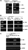
The carboxy terminus of Caprin-1 selectively binds mRNAs associated with cellular proliferation through a mechanism dependent on the RGG motifs. (A) mRNA for c-Myc and cyclin D2 coprecipitate with endogenous Caprin-1 and G3BP-1. 293T cells were lysed in polysome lysis buffer. The supernatants of these lysates were precleared as described using beads coated with normal rabbit serum or mouse IgG1 for the anti-Caprin-1 and anti-G3BP-1 precipitations, respectively. Precleared polysome lysates were then incubated with protein A-Sepharose beads, which had been conjugated with anti-Caprin-1 rabbit serum, control rabbit serum, or protein G-Sepharose beads conjugated with the anti-G3BP-1 mouse MAb or an isotype-matched control mouse IgG1 MAb. RNA was extracted from the proteins bound to the beads after IP, as described above. RT-PCR was performed as described to detect the presence of c-Myc, cyclin D2, and GAPDH transcripts. (B) A carboxy-terminal fragment of Caprin-1 (381 to 709) that fails to bind G3BP-1 selectively binds mRNA for c-Myc and cyclin D2. As before, 293T cells were transfected with the indicated Flag-tagged fragments comprising the carboxy termini of Caprin-1 (381 to 709) or G3BP-1 (142 to 466) or the amino-terminal HR1 region of Caprin-1, Caprin-1 (47 to 380). The fragments were immunoprecipitated with anti-Flag as described above, and associated mRNA were assayed as before. Analysis of IP by SDS-PAGE and immunoblotting with antibody to Caprin-1 and G3BP-1 confirmed that fragments did not interact with endogenous protein (data not shown). (C) The RGG motifs in Caprin-1 are essential for selective binding of c-Myc and cyclin D2 mRNA. As before, 293T cells were transfected with a Flag-tagged carboxy-terminal fragment of Caprin-1 (381 to 709), a fragment in which each of the RGG motif had been mutated to AGG (AGGX3), or a Caprin-1 fragment that was truncated at residue 606 so it lacked the RGG motifs entirely, Caprin-1 (381to 605). The fragments were immunoprecipitated with anti-Flag and associated mRNAs were assayed as before.
FIG. 11.
FIG. 11.
Global protein synthesis is inhibited by overexpression of Caprin-1 but is not affected by the absence of Caprin-1. (A) Overexpression of Caprin-1 inhibits protein synthesis. HeLa cells transiently expressing GFP alone or GFP-Caprin-1 were purified using a fluorescence-activated cell sorter and assessed for the rates of protein synthesis by incorporation of [3H]leucine as described above. As a positive control for inhibition of protein synthesis, nontransfected cells were treated with 10 μg/ml CHX. Note the significant decrease in global rates of protein synthesis in cells expressing GFP-Caprin-1 compared with those expressing equimolar amounts of GFP alone. (B) Cells lacking Caprin-1 exhibit no global changes in protein synthesis. Avian R-Caprin-1−/− DT40 cells were grown in the presence of DOX for 3 days to suppress the expression of human Caprin-1 to undetectable levels. Together with control R-Caprin-1−/− cells cultured in the absence of DOX, they were assessed for rates of protein synthesis by incorporation of radioactive leucine label as described. Parental DT40 cells were treated with CHX (10 μg/ml) as a positive control. Note that the R-Caprin−/− DT40 exhibited similar rates of protein synthesis in the presence (+) or absence (−) of DOX. Results are expressed as means ± standard errors of the means.

References

    1. Angenstein, F., A. M. Evans, S. C. Ling, R. E. Settlage, S. Ficarro, F. A. Carrero-Martinez, J. Shabanowitz, D. F. Hunt, and W. T. Greenough. 2005. Proteomic characterization of messenger ribonucleoprotein complexes bound to nontranslated or translated poly(A) mRNAs in the rat cerebral cortex. J. Biol. Chem. 280:6496-6503. - PubMed
    1. Angenstein, F., A. M. Evans, R. E. Settlage, S. T. Moran, S. C. Ling, A. Y. Klintsova, J. Shabanowitz, D. F. Hunt, and W. T. Greenough. 2002. A receptor for activated C kinase is part of messenger ribonucleoprotein complexes associated with polyA-mRNAs in neurons. J. Neurosci. 22:8827-8837. - PMC - PubMed
    1. Atlas, R., L. Behar, E. Elliott, and I. Ginzburg. 2004. The insulin-like growth factor mRNA binding-protein IMP-1 and the Ras-regulatory protein G3BP associate with tau mRNA and HuD protein in differentiated P19 neuronal cells. J. Neurochem. 89:613-626. - PubMed
    1. Bassell, G. J., C. M. Powers, K. L. Taneja, and R. H. Singer. 1994. Single mRNAs visualized by ultrastructural in situ hybridization are principally localized at actin filament intersections in fibroblasts. J. Cell Biol. 126:863-876. - PMC - PubMed
    1. Ben-Asouli, Y., Y. Banai, Y. Pel-Or, A. Shir, and R. Kaempfer. 2002. Human interferon-gamma mRNA autoregulates its translation through a pseudoknot that activates the interferon-inducible protein kinase PKR. Cell 108:221-232. - PubMed

Publication types

MeSH terms