. 2007 Mar;27(5):1881-8.
doi: 10.1128/MCB.01122-06. Epub 2007 Jan 8.

Hepatic de novo lipogenesis is present in liver-specific ACC1-deficient mice

Affiliations

Hepatic de novo lipogenesis is present in liver-specific ACC1-deficient mice

Naomoto Harada et al. Mol Cell Biol. 2007 Mar.

Erratum in

  • Mol Cell Biol. 2007 May;27(9):3556

Abstract

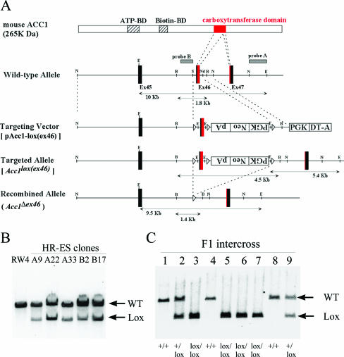
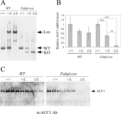
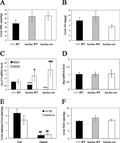
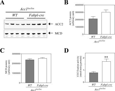
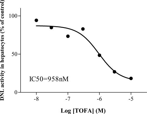
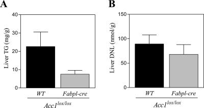
Acetyl coenzyme A (acetyl-CoA) carboxylase (ACC) catalyzes carboxylation of acetyl-CoA to form malonyl-CoA. In mammals, two isozymes exist with distinct physiological roles: cytosolic ACC1 participates in de novo lipogenesis (DNL), and mitochondrial ACC2 is involved in negative regulation of mitochondrial beta-oxidation. Since systemic ACC1 null mice were embryonic lethal, to clarify the physiological role of ACC1 in hepatic DNL, we generated the liver-specific ACC1 null mouse by crossbreeding of an Acc1(lox(ex46)) mouse, in which exon 46 of Acc1 was flanked by two loxP sequences and the liver-specific Cre transgenic mouse. In liver-specific ACC1 null mice, neither hepatic Acc1 mRNA nor protein was detected. However, to compensate for ACC1 function, hepatic ACC2 protein and activity were induced 1.4 and 2.2 times, respectively. Surprisingly, hepatic DNL and malonyl-CoA were maintained at the same physiological levels as in wild-type mice. Furthermore, hepatic DNL was completely inhibited by an ACC1/2 dual inhibitor, 5-tetradecyloxyl-2-furancarboxylic acid. These results strongly demonstrate that malonyl-CoA from ACC2 can access fatty acid synthase and become the substrate for the DNL pathway under the unphysiological circumstances that result with ACC1 disruption. Therefore, there does not appear to be strict compartmentalization of malonyl-CoA from either of the ACC isozymes in the liver.


Figures

FIG. 1.
FIG. 1.
Conditional deletion of a carboxyltransferase domain of the Acc1 gene by the Cre/loxP system. A. Schematic of the targeting strategy. The structures are shown for ACC1 protein, the wild-type allele, the targeting vector pAcc1-lox(ex46), targeted allele Acc1lox(ex46), and the Cre-recombined allele (Acc1Δex46). The ATP- and biotin-binding domains (ATP-BD and Biotin-BD) are shown as striped boxes, whereas the carboxyltransferase domain in exons 46 and 47 is shown as a red box. The exon numbers are shown as the protein-coding exon numbers. The PGK-neo-bpA and PGK-DT-a cassettes are shown as open boxes. The positions of Southern hybridization probes A and B, which detect EcoRI and BglII fragments, respectively, are also shown. Restriction enzyme sites are the following: B, BamHI; E, EcoRI; N, NcoI; Nd, NdeI; and S, SalI. B. Southern blot analysis of homologous recombination in the targeted ES cell clones. The EcoRI-digested DNA fragments from the parental ES cell line RW4 and targeted ES clones (A9, A22, A33, B2, and B17) were hybridized with probe A. Arrows indicate wild-type (WT; 10 kb) and targeted (Lox; 5.4 kb) alleles. C. Southern blot analysis of the intercross offspring of Acc1+/lox(ex46) heterozygotes. The EcoRI-digested tail DNA from each mouse was hybridized with probe A. The determined genotypes are shown on the bottom.
FIG. 2.
FIG. 2.
Conditional KO of the Acc1 gene in liver. A. Southern blot analysis of the liver DNA from the wild-type (+/+), Acc1+/lox(ex46) (+/L), and Acc1lox(ex46)/lox(ex46) (L/L) mice with or without the Fabpl-cre transgenic allele. The BglII-digested DNA fragments were hybridized with probe B. Arrows indicate targeted (Lox; 4.5 kb), wild-type (WT; 1.8 kb), and recombined (KO; 1.4 kb) alleles. B. Relative amount of hepatic Acc1 mRNA in each genotype (n = 3). The Acc1 expression level was measured by quantitative real-time PCR analysis, normalized with the endogenous β-actin mRNA level, and is shown as an amount relative to that in the wild-type mice. **, P < 0.01; ***, P < 0.001. C. Western blot analysis of hepatic ACC1 protein. Total protein was loaded on gradient (3% to 8%) polyacrylamide gel electrophoresis gel (50 μg/lane) and detected with anti-ACC1 antibody (Ab).
FIG. 3.
FIG. 3.
Key parameters related to the DNL pathway in the liver. (A) DNL and (B) TG levels in the livers obtained from Acc1+/+ WT (+/+:WT; black column; n = 10), Acc1lox(ex46)/lox(ex46) WT (lox/lox:WT; gray column; n = 9), and Acc1lox(ex46)/lox(ex46) TgFabpl-cre (lox/lox:cre; open column; n = 10) mice. (C) Relative Acc1 (black column) and Acc2 (open column) and (D) Mcd mRNA levels in the livers obtained from +/+:WT (n = 10), lox/lox:WT (n = 9), and lox/lox:cre (n = 10) mice. ***, P < 0.001 compared with +/+:WT (for Acc1); #, P < 0.05; ###, P < 0.001 compared with +/+:WT (for Acc2). (E) Malonyl-CoA level in the livers obtained from +/+:WT (black column; n = 5) and lox/lox:cre (open column; n = 6) mice fed with a normal chow diet or fasted for 16 h. ##, P < 0.01 for fed versus fasted groups within the same genotype. (F) FAO in the livers obtained from +/+:WT (black column; n = 10), lox/lox:WT (gray column; n = 9), and lox/lox:cre (open column; n = 11) mice. Each column represents average results ± standard errors.
FIG. 4.
FIG. 4.
ACC2 and MCD levels in the liver from Acc1-lox and Acc1 cKO mice. (A) Immunoblot images of ACC2 and MCD proteins. Quantification of immunoblot images of ACC2 (B) and MCD (C) proteins in the liver from Acc1-lox (gray column; n = 4) and Acc1 cKO (open column; n = 4). (D) ACC2 activity in the liver from Acc1-lox (gray column; n = 4) and Acc1 cKO (open column; n = 4). Each column represents average results ± standard errors. **, P < 0.01.
FIG. 5.
FIG. 5.
Inhibitory effect of TOFA on DNL in primary hepatocytes isolated from Acc1 cKO mice. Primary hepatocytes prepared from Acc1 cKO mice were incubated with TOFA (10 nM ∼ 10 μM). Each symbol represents the average of two or three triplicate experiments and is shown as a percentage of control DNL activity.
FIG. 6.
FIG. 6.
Liver TG (A) and DNL (B) in Acc1-lox and Acc1 cKO mice on HSD. Acc1-lox (black column; n = 6) and Acc1 cKO mice (gray column; n = 4) were fed with HSD for 3 days. Each column represents average results ± standard errors.

References

    1. Abu-Elheiga, L., D. B. Almarza-Ortega, A. Baldini, and S. J. Wakil. 1997. Human acetyl-CoA carboxylase 2. Molecular cloning, characterization, chromosomal mapping, and evidence for two isoforms. J. Biol. Chem. 272:10669-10677. - PubMed
    1. Abu-Elheiga, L., W. R. Brinkley, L. Zhong, S. S. Chirala, G. Woldegiorgis, and S. J. Wakil. 2000. The subcellular localization of acetyl-CoA carboxylase 2. Proc. Natl. Acad. Sci. USA 97:1444-1449. - PMC - PubMed
    1. Abu-Elheiga, L., A. Jayakumar, A. Baldini, S. S. Chirala, and S. J. Wakil. 1995. Human acetyl-CoA carboxylase: characterization, molecular cloning, and evidence for two isoforms. Proc. Natl. Acad. Sci. USA 92:4011-4015. - PMC - PubMed
    1. Abu-Elheiga, L., M. M. Matzuk, K. A. Abo-Hashema, and S. J. Wakil. 2001. Continuous fatty acid oxidation and reduced fat storage in mice lacking acetyl-CoA carboxylase 2. Science 291:2613-2616. - PubMed
    1. Abu-Elheiga, L., M. M. Matzuk, P. Kordari, W. Oh, T. Shaikenov, Z. Gu, and S. J. Wakil. 2005. Mutant mice lacking acetyl-CoA carboxylase 1 are embryonically lethal. Proc. Natl. Acad. Sci. USA 102:12011-12016. - PMC - PubMed

Publication types