. 2007 Mar;27(6):2130-43.
doi: 10.1128/MCB.01826-06. Epub 2007 Jan 8.

A triple helix within a pseudoknot is a conserved and essential element of telomerase RNA

Affiliations

A triple helix within a pseudoknot is a conserved and essential element of telomerase RNA

Kinneret Shefer et al. Mol Cell Biol. 2007 Mar.

Abstract

Telomerase copies a short template within its integral telomerase RNA onto eukaryotic chromosome ends, compensating for incomplete replication and degradation. Telomerase action extends the proliferative potential of cells, and thus it is implicated in cancer and aging. Nontemplate regions of telomerase RNA are also crucial for telomerase function. However, they are highly divergent in sequence among species, and their roles are largely unclear. Using in silico three-dimensional modeling, constrained by mutational analysis, we propose a three-dimensional model for a pseudoknot in telomerase RNA of the budding yeast Kluyveromyces lactis. Interestingly, this structure includes a U-A.U major-groove triple helix. We confirmed the triple-helix formation in vitro using oligoribonucleotides and showed that it is essential for telomerase function in vivo. While triplex-disrupting mutations abolished telomerase function, triple compensatory mutations that formed pH-dependent G-C.C(+) triples restored the pseudoknot structure in a pH-dependent manner and partly restored telomerase function in vivo. In addition, we identified a novel type of triple helix that is formed by G-C.U triples, which also partly restored the pseudoknot structure and function. We propose that this unusual structure, so far found only in telomerase RNA, provides an essential and conserved telomerase-specific function.


Figures
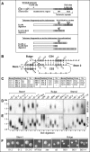
FIG. 1.
FIG. 1.
Mutational analysis of the pseudoknot element. (A) Schematic representation showing telomeric fragments digested with restriction endonucleases EcoRI or EcoRI plus BclI and hybridization probes (WT and BclI specific). (B and C) Secondary structure model of the K. lactis pseudoknot element (B), with the positions of the mutations introduced to test stem 1, bulge, and stem 2 (C) highlighted in gray. (D and E) Plasmids carrying ter1 genes with the template BclI mutation alone (WT-BclI), additional pseudoknot mutations, or an empty vector (Δter), as indicated above the lanes (D), were used to replace the WT TER1 gene encoded on a URA plasmid. Genomic DNA was prepared from the resulting K. lactis strains in their sixth passage and analyzed by a Southern blot hybridized first with a BclI-specific (D) and then with a WT (E) probe. Only portions of the gel are shown, which include a group of 7 out of the 12 K. lactis telomeres. (F) Typical colony phenotypes of K. lactis strains taken at their fourth passage. Impaired telomere maintenance is associated with rough colony appearance, as opposed to the smooth WT colonies.
FIG. 2.
FIG. 2.
Mutational analysis of the triple-helical stem. (A) Secondary structure model of the K. lactis pseudoknot element. Highlighted in gray are the positions of the mutations introduced into the TER1 gene to test the triple-helical stem. (B and C) The mutations are described (B) and indicated in gray on the small schematic representations of the pseudoknot above the lanes in panel C. (C and D) Genomic DNA was prepared from the various K. lactis strains in their sixth passage and analyzed with a Southern blot hybridized with Bcl-specific (C) and WT (D) probes, as described for Fig. 1. (E) Typical colony phenotypes of the strains at their fourth passage. (F) WT hybridization to telomere fragments of strains grown at 16°C (for six passages on plates and then in liquid medium) instead of at the normal temperature of 28 to 30°C.
FIG. 3.
FIG. 3.
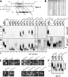
Dimer formation by oligoribonucleotides. (A and B) Secondary structure model of the K. lactis pseudoknot element (A), with the positions of the mutations introduced to test stem 2 and the CS3 loop (B) highlighted in gray. The first G residue in CS4 (in lowercase) is not present in the WT sequence but was introduced to facilitate efficient transcription by T7 RNA polymerase. (C) The WT and mutated CS3 and CS4 oligoribonucleotides (0.01 pmol each) described in panels A and B, both radioactively labeled, were denatured, renatured, and analyzed by 15% nondenaturing PAGE at 4°C and pH 6.25. Single oligoribonucleotides are indicated by CS3 or CS4 and asterisks below the lanes. Lanes with mixtures of both CS3 and CS4 are labeled by the name (above) and number (below) of the mutations introduced. Note that the mutant CS4-S2.3 oligoribonucleotide migrated slower than the WT CS4, presumably because the mutated (but not the WT) CS4 alone can form a stable dimer. Since it comigrated with the longer CS3, only one band corresponding to the monomer form appears under S2.3 and S2.3+3′.
FIG. 4.
FIG. 4.
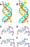
Triple-helix models. Modeling was performed using miniCarlo as described in Materials and Methods. (A and B) Eleven triples are shown for poly(rU)-poly(rA) · poly(rU) (A) and poly(rC)-poly(rG) ·poly(rU) (B). In each strand, conformations of consecutive residues are identical. Watson-Crick strands are colored in brown (purine strand) and yellow (pyrimidine strand), and the third poly(rU) strand is colored in cyan. Backbones are schematically shown as ribbons, and sugar O4′ atoms are colored in red. (C) A typical U-A·U triple from the WT pseudoknot model. (D) A C-G·U base triple from the model of the pseudoknot structure for the S2.3+3′ mutant. (E) An example of a single C-G·U base triple found in the crystal structure of the Haloarcola marismortui 50S ribosomal subunit (1). (F) An example of a C-G·C+ base triple from a DNA triplex (PDB 149D) (23).
FIG. 5.
FIG. 5.
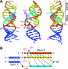
A three-dimensional model for the wild-type pseudoknot structure. Modeling was performed using miniCarlo as described in Materials and Methods. (A) A ribbon representation (three different views). Stem 1 is shown in blue, Watson-Crick strands of stem 2 are colored in brown (purine-rich strand) and yellow (pyrimidine-rich strand), and the third oligo(U) strand is colored in cyan. Bulged-out U959 is shown in magenta; unpaired residues C861, C877, and C957 underneath the last triple are in red; and the Watson-Crick pair C878-G956 terminating stem 2 are in gray. (B) A schematic representation of base-pairing in the pseudoknot. Vertical lines represent Watson-Crick interactions; tilted lines, Hoogsteen interactions; and the asterisk, a G·U wobble pair. Note that if protonated, C861 has a potential to form a base triple (C878-G956·C861+).
FIG. 6.
FIG. 6.
pH-dependent dimer formation by oligoribonucleotides. (A and B) A schematic model of the K. lactis pseudoknot with five base triples (A), with the positions of the mutations (B) highlighted in gray. (C and D) Radioactively labeled CS4 and unlabeled CS3 (0.1 and 0.2 pmol, respectively) were analyzed as described in the legend to Fig. 3 and Materials and Methods. Identical gels were run simultaneously at pH 5 (C) and pH 7 (D). Single oligoribonucleotides are indicated by CS4 above and asterisks below the lanes. Lanes with mixtures of both CS3 and CS4 are indicated by the name (above) and number (below) of mutations introduced. (E) UV melting experiments were performed using various oligoribonucleotides at pH 5, 6, and 7. The Tm values were determined as described in Materials and Methods.
FIG. 7.
FIG. 7.
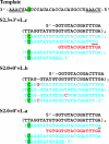
Incorporation of truncated and mutated repeats onto telomeres in vivo. Shown are examples of cloned telomere sequences from the S2.3+3′+L.c, S2.0+0′+Lb, and S2.0+0′+La mutant strains at their sixth passage after introducing the mutant ter gene. The telomeric repeats are aligned to the template sequence above. Underlined are the repeated sequence at the beginning and the end of the template. Underlined in the S2.3+3′+L.c and S2.0+0′+La telomeres are nucleotides used for base-pairing and initiating the synthesis of truncated repeats from the middle of the template. The telomerase template and wild-type telomeric repeats are shown in black; BclI repeats synthesized by the mutant telomerase, in blue; truncated repeats and misincorporated nucleotides, in red. The BclI mutations are highlighted in green.
FIG. 8.
FIG. 8.
Conservation of the triple-helix pseudoknot in the “Kluyveromyces marxianus” cluster of species. Pseudoknot models are shown for the TER sequences of six Kluyveromyces species (32). Conserved residues are highlighted in gray. Hoogsteen interactions are indicated as tilted lines.

Similar articles

Cited by

References

    1. Ban, N., P. Nissen, J. Hansen, P. B. Moore, and T. A. Steitz. 2000. The complete atomic structure of the large ribosomal subunit at 2.4 A resolution. Science 289:905-920. - PubMed
    1. Cech, T. R. 2004. Beginning to understand the end of the chromosome. Cell 116:273-279. - PubMed
    1. Chappell, A. S., and V. Lundblad. 2004. Structural elements required for association of the Saccharomyces cerevisiae telomerase RNA with the Est2 reverse transcriptase. Mol. Cell. Biol. 24:7720-7736. - PMC - PubMed
    1. Chen, J. L., M. A. Blasco, and C. W. Greider. 2000. Secondary structure of vertebrate telomerase RNA. Cell 100:503-514. - PubMed
    1. Church, G. M., and W. Gilbert. 1984. Genomic sequencing. Proc. Natl. Acad. Sci. USA 81:1991-1995. - PMC - PubMed

Publication types

LinkOut - more resources