

Rat model of polymicrobial infection, immunity, and alveolar bone resorption in periodontal disease
- PMID: 17210663
- PMCID: PMC1865722
- DOI: 10.1128/IAI.00733-06
Rat model of polymicrobial infection, immunity, and alveolar bone resorption in periodontal disease
Abstract
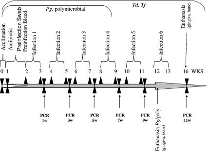
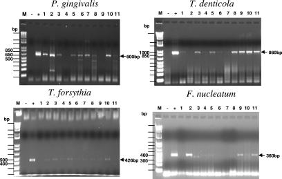
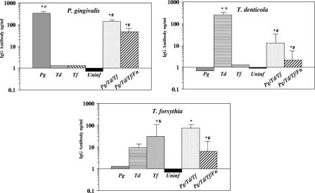
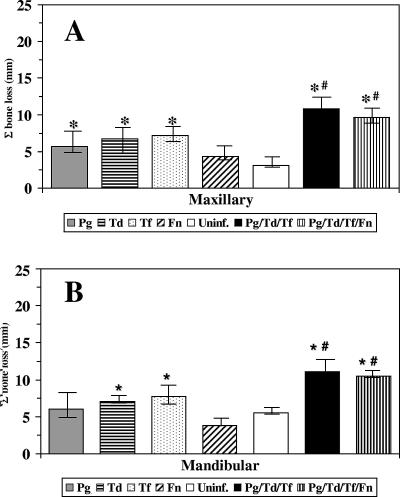
One of the predominant polymicrobial infections of humans is expressed clinically as periodontal disease. Porphyromonas gingivalis, Treponema denticola, and Tannerella forsythia have been strongly implicated as members of a pathogenic consortium in the etiology of adult periodontitis. In this study we hypothesized that P. gingivalis, T. denticola, and T. forsythia are synergistic in terms of virulence potential and induce chronic periodontal inflammation that leads to alveolar bone resorption in a polymicrobial infection in rats. Groups of rats were infected with either P. gingivalis, T. denticola, or T. forsythia in monomicrobial infections or with all three species in polymicrobial oral infections with or without Fusobacterium nucleatum. PCR analyses of oral microbial samples demonstrated that rats infected with one bacterium were orally colonized by each of the bacteria during the study interval, and increased serum immunoglobulin G (IgG) antibody levels substantiated the interaction of the host with the infecting bacteria. PCR analyses of the rats with polymicrobial infections demonstrated that most rats were infected with P. gingivalis, T. denticola, and T. forsythia as a consortium. Furthermore, all rats exhibited a significant increase in the level of IgG antibody to the polymicrobial consortium. Radiographic measurement of alveolar bone resorption showed that rats infected with the polymicrobial consortium with or without F. nucleatum exhibited significantly increased alveolar bone resorption compared to the resorption in uninfected control rats, as well as the resorption in rats infected with one of the microbes. These results documented that P. gingivalis, T. denticola, and T. forsythia not only exist as a consortium that is associated with chronic periodontitis but also exhibit synergistic virulence resulting in the immunoinflammatory bone resorption characteristic of periodontitis.
Figures
Similar articles
-
Bis-enoxacin blocks rat alveolar bone resorption from experimental periodontitis.PLoS One. 2014 Mar 17;9(3):e92119. doi: 10.1371/journal.pone.0092119. eCollection 2014. PLoS One. 2014. PMID: 24638087 Free PMC article.
-
Periodontal pathogens invade gingiva and aortic adventitia and elicit inflammasome activation in αvβ6 integrin-deficient mice.Infect Immun. 2015 Dec;83(12):4582-93. doi: 10.1128/IAI.01077-15. Epub 2015 Sep 14. Infect Immun. 2015. PMID: 26371120 Free PMC article.
-
Mouse model of experimental periodontitis induced by Porphyromonas gingivalis/Fusobacterium nucleatum infection: bone loss and host response.J Clin Periodontol. 2009 May;36(5):406-10. doi: 10.1111/j.1600-051X.2009.01393.x. J Clin Periodontol. 2009. PMID: 19419440
-
[Pathogenic potential of Porphyromonas gingivalis, Treponema denticola and Tannerella forsythia, the red bacterial complex associated with periodontitis].Pathol Biol (Paris). 2007 Apr-May;55(3-4):154-62. doi: 10.1016/j.patbio.2006.07.045. Epub 2006 Oct 17. Pathol Biol (Paris). 2007. PMID: 17049750 Review. French.
-
Porphyromonas gingivalis: major periodontopathic pathogen overview.J Immunol Res. 2014;2014:476068. doi: 10.1155/2014/476068. Epub 2014 Mar 25. J Immunol Res. 2014. PMID: 24741603 Free PMC article. Review.
Cited by
-
Impact of Silicon Carbide Coating and Nanotube Diameter on the Antibacterial Properties of Nanostructured Titanium Surfaces.Materials (Basel). 2024 Aug 2;17(15):3843. doi: 10.3390/ma17153843. Materials (Basel). 2024. PMID: 39124507 Free PMC article.
-
Kanamycin Resistance Cassette for Genetic Manipulation of Treponema denticola.Appl Environ Microbiol. 2015 Jul;81(13):4329-38. doi: 10.1128/AEM.00478-15. Epub 2015 Apr 17. Appl Environ Microbiol. 2015. PMID: 25888173 Free PMC article.
-
Subgingival biofilm formation.Periodontol 2000. 2010 Feb;52(1):38-52. doi: 10.1111/j.1600-0757.2009.00311.x. Periodontol 2000. 2010. PMID: 20017794 Free PMC article. Review. No abstract available.
-
Persistence of Tannerella forsythia and Fusobacterium nucleatum in dental plaque: a strategic alliance.Curr Oral Health Rep. 2020 Mar;7(1):22-28. doi: 10.1007/s40496-020-00254-6. Epub 2020 Jan 29. Curr Oral Health Rep. 2020. PMID: 36779221 Free PMC article.
-
Bis-enoxacin blocks rat alveolar bone resorption from experimental periodontitis.PLoS One. 2014 Mar 17;9(3):e92119. doi: 10.1371/journal.pone.0092119. eCollection 2014. PLoS One. 2014. PMID: 24638087 Free PMC article.
References
-
- Baker, P. J., R. T. Evans, and D. C. Roopenian. 1994. Oral infection with Porphyromonas gingivalis and induced alveolar bone loss in immunocompetent and severe combined immunodeficient mice. Arch. Oral Biol. 39:1035-1040. - PubMed
-
- Brogden, K. A. 2002. Polymicrobial diseases of animals and humans, p. 3-20. In K. Brogden and J. Guthmiller (ed.), Polymicrobial diseases. ASM Press, Washington, DC. - PubMed
Publication types
MeSH terms
Substances
Grants and funding
LinkOut - more resources
Full Text Sources
Other Literature Sources
Molecular Biology Databases
