. 2007 Jan 29;96(2):269-76.
doi: 10.1038/sj.bjc.6603553. Epub 2007 Jan 9.

Potentiation of antitumour activity of docetaxel by combination with trastuzumab in a human prostate cancer xenograft model and underlying mechanisms

Affiliations

Potentiation of antitumour activity of docetaxel by combination with trastuzumab in a human prostate cancer xenograft model and underlying mechanisms

M-E Legrier et al. Br J Cancer. .

Abstract

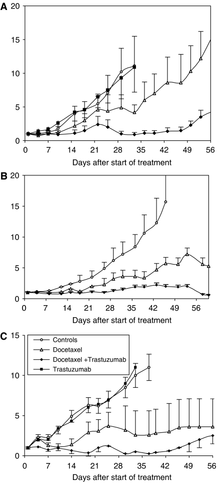
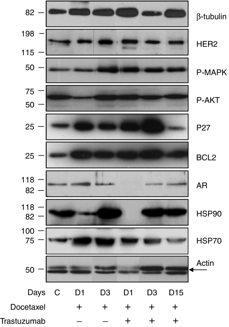
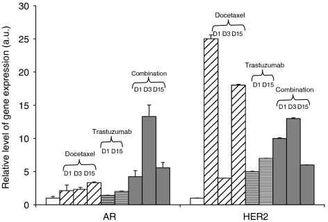
Antitumour activity of docetaxel (Taxotere) in hormone-dependent (HD) and hormone-independent (HID) prostate cancer PAC120 xenograft model was previously reported, and its level was associated with HER2 protein expression. In the present study, we evaluate the antitumour effects of docetaxel combined with trastuzumab (Herceptin), an anti-HER2 antibody. Although trastuzumab alone had no effect on tumour growth, it potentiated the antitumour activity of docetaxel in HD tumours and more strongly in HID variants. Using the HID28 variant, we show that docetaxel treatment of tumour-bearing mice induces an increased HER2 mRNA expression of the tyrosine kinase receptor of 25-fold 24 h after docetaxel treatment, while HER2 protein and p-AKT decreased. This was followed by an increase of HER2 protein 3 days (two-fold) after docetaxel treatment and by a strong HER2 release in the serum of treated mice; expression of phospho-ERK, p27, BCL2 and HSP70 concomitantly increased. Similar molecular alterations were induced by docetaxel plus trastuzumab combination, except for that there was a transient and complete disappearance of AR and HSP90 proteins 24 h after treatment. We show that in addition to its known effects on tubulin and mitotic spindles, docetaxel induces complex signalisation pathway mechanisms in surviving cells, including HER2, which can be pharmacologically targeted. This study suggests that the docetaxel/trastuzumab combination may prove an effective therapeutic approach for HER2-expressing hormone-refractory prostate cancer.


Figures

Figure 1
Figure 1
Effect of docetaxel and trastuzumab, alone or combined, on the growth of PAC120, HID25 and HID28 prostate cancer xenografts. Tumour growth curves in mice that were untreated (empty circles), treated with docetaxel, 20 mg kg−1 every 3 weeks (empty triangles), trastuzumab, 10 mg kg−1 weekly (filled squares) and docetaxel plus trastuzumab (filled lozenges). (A) Growth curves of the PAC120 HID prostate cancer xenograft. (B) Growth curves of the HID25 HID prostate cancer xenograft grown in castrated mice. (C) Growth curves of the HID28 HID prostate cancer xenograft grown in castrated mice. Bars represent s. d.
Figure 2
Figure 2
Histological features of PAC120, HID25 and HID28 xenografts (HES staining, × 200 magnification). Left panels, untreated tumours; middle panels, docetaxel-treated tumours; right panels, docetaxel plus trastuzumab-treated tumours. (A) PAC120 tumours were characterised by a Gleason score of 9 (5+4) with compact sheets of tumour cells surrounded by a fine murine stroma. (B) At 3 days after docetaxel treatment, accumulation of mitotic cells is visible (arrows). (C) At 47 days after treatment with docetaxel plus trastuzumab, the tumour has markedly regressed, leaving large areas of empty fibro-adenomatous tumour stroma (arrow), invaded by murine cells. (D) HID25 tumours display mucinous differentiation with principally signet-ring cells. (E) At 3 days after docetaxel treatment, accumulation of numerous and abnormal mitotic cells (arrows) (HES × 400). (F) At 3 days after treatment with the docetaxel plus trastuzumab combination, presence of elongated tumour cells with hyperchromatic nuclei (arrows). (G) HID28 shows histologic features similar to that of PAC120. (H) At 3 days after docetaxel treatment, there is accumulation of mitotic cells, multipolar divisions and few apoptotic bodies (arrows). (I) At 3 days after treatment with the docetaxel plus trastuzumab combination, note the presence of enlarged and mitotic tumour cells in the middle of compact cellular masses, surrounded by elongated cells (arrows). Percent indicates the mitotic index.
Figure 3
Figure 3
Expression pattern of HER2 and HSP90 in HID28 xenografts by immunohistochemistry. Left two panels, untreated tumours; middle two panels, docetaxel-treated tumours at day 3; right two panels, docetaxel/trastuzumab treatment (× 200 magnification). HER2 immunodetection. (A) The anti-HER2 antibody staining is weak, diffuse and focal, stronger in mitotic cells (arrows). (B) HER2 staining is increased in all the tumour cells with preferential membrane staining. (C) Immunostaining was restricted to the membrane of mitotic cells at day 15. HSP90 immunodetection. (D) Anti-HSP90 antibody stained the cytoplasm of HID28 tumour cells. (E) All tumour cells are stained except mitotic cells (arrow). (F) The anti-HSP90 staining disappeared from all tumour cells, apart from rare cells at day 1.
Figure 4
Figure 4
Western blotting analysis of protein expression in a representative HID28 xenograft, after one cycle of treatment. Untreated tumours (lane 1), 1 or 3 days after docetaxel treatment (lanes 2 and 3, respectively); 1, 3 or 15 days after docetaxel plus trastuzumab treatment (lanes 4, 5 and 6, respectively). Actin (45 kDa) was used as a control. Signals correspond to β-tubulin (110 kDa), HER2 (185 kDa), phospho-ERK (42–44 kDa), phospho-AKT (60 kDa), p27 (27 kDa), BCL2 (26 kDa) AR (110 kDa), HSP90 (90 kDa) and HSP70 (70kDa). Histograms of proteic expression of HER2, AR and HSP90 in treated tumours reported to that of controls, day 1, 3 or 15 after docetaxel treatment alone or combined with trastuzumab.
Figure 5
Figure 5
Real-time RT–PCR quantitation of AR and HER2 mRNA transcripts in HID28 xenograft, after one cycle of treatment. Median levels of five tumours for each treatment group. Gene expression, performed in triplicate, was normalised against the HPRT gene and expressed relative to the control group. Bars represent s.d.

References

    1. Abal M, Andreu JM, Barasoain I (2003) Taxanes: microtubule and centrosome targets, and cell cycle dependent mechanisms of action. Curr Cancer Drug Targets 3: 193–203 - PubMed
    1. Agus DB, Scher HI, Higgins B, Fox WD, Heller G, Fazzari M, Cordon-Cardo C, Golde DW (1999) Response of prostate cancer to anti-Her-2/neu antibody in androgen-dependent and -independent human xenograft models. Cancer Res 59: 4761–4764 - PubMed
    1. Burns RG, Islam K, Chapman R (1984) The multiple phosphorylation of the microtubule-associated protein MAP2 controls the MAP2:tubulin interaction. Eur J Biochem 141: 609–615 - PubMed
    1. Calvo BF, Levine AM, Marcos M, Collins QF, Iacocca MV, Caskey LS, Gregory CW, Lin Y, Whang YE, Earp HS, Mohler JL (2003) Human epidermal receptor-2 expression in prostate cancer. Clin Cancer Res 9: 1087–1097 - PubMed
    1. Craft N, Shostak Y, Carey M, Sawyers CL (1999) A mechanism for hormone-independent prostate cancer through modulation of androgen receptor signaling by the HER-2/neu tyrosine kinase. Nat Med 5: 280–285 - PubMed

Publication types

MeSH terms