

doi: 10.1021/nl0626434.
Semiconductor quantum rods as single molecule fluorescent biological labels
Affiliations
- PMID: 17212460
- PMCID: PMC3984543
- DOI: 10.1021/nl0626434
Item in Clipboard
Semiconductor quantum rods as single molecule fluorescent biological labels
Nano Lett.
2007 Jan.
Abstract
In this paper, we report the development of rod-shaped semiconductor nanocrystals (quantum rods) as fluorescent biological labels. Water-soluble biocompatible quantum rods have been prepared by surface silanization and applied for nonspecific cell tracking as well as specific cellular targeting. Quantum rods are brighter single molecule probes as compared to quantum dots. They have many potential applications as biological labels in situations where their properties offer advantages over quantum dots.
Figures
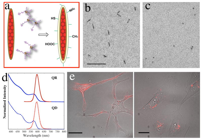
(a) A cartoon illustrating silanization of quantum rods. Crosslinked silanes are priming molecules for the surface coating. (b) TEM image of silanized rods in neutral phosphate buffer. Scale bar = 100 nm. (c) TEM image of silanized dots in neutral phosphate buffer. Scale bar = 100 nm. (d) The UV-Vis absorption and emission spectra of silanized QR and QD. The blue curves are the absorption spectra; the red curves are the emission spectra. (e) Silanized QRs are biocompatible and non-toxic to living cells. The red fluorescence in the images is from QRs in human breasts cancer cells MDA-MB-231 after 1h (left) and 24h (right) transfected with Chariot™. These are merged images of transmission and fluorescent micrograms. Scale bar is 20 μm.
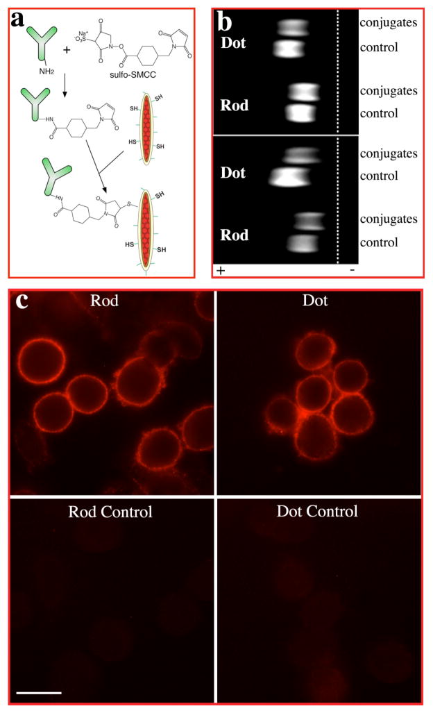
(a) Scheme for antibody bioconjugation of quantum rods. (b) Electrophoresis analyses of quantum rods/dots bioconjugation. Top, quantum rods/dots conjugated with F(ab′)2 fragment of goat anti-mouse IgG antibody. Bottom, quantum rods/dots conjugated with whole goat anti-mouse IgG antibody. The conjugates moved slower than the free nanocrystals (control) due to the linkage with antibodies. (c) Immunofluorescence labeling of breast cancer cell marker Her2 on breast cancer cells SK-BR-3. The Her2 marker was labeled with mouse anti-Her2 antibody and goat anti-mouse IgG F(ab′)2 conjugated quantum rods/dots. The bottom images show that there is minimum binding of free nanocrystals to the anti-Her2 antibody treated cells. Scale bar is 20 μm.
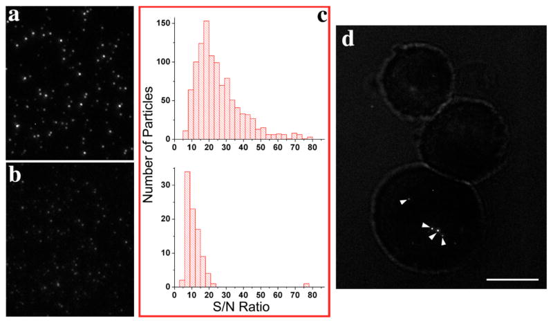
Fluorescence microscope images show that at the single molecule level, QRs (a) are much brighter than QDs (b). (c) Statistical results of S/N distribution of QRs (top) and QDs (bottom) from 15 image sequences. The mean S/N for single rods is 26, while it is 11 for single dots. (d) Single QRs (indicated by arrows) are still very bright inside live MDA-MB-231 human breast cancer cells. Scale bar is 10 μm.
References
-
- Nam J, Thaxton CS, Mirkin CA. Science. 2003;301:1884–1886. - PubMed
-
- Sonnichsen C, Reinhard BM, Liphardt J, Alivisatos AP. Nature Biotechnology. 2005;23(6):741–745. - PubMed
-
- Turner JL, Pan D, Plummer R, Chen Z, Whittaker AK, Wooley KL. Advanced Functional Materials. 2005;15(8):1248–1254.
-
- Jun YW, HYM, Choi JS, Lee JH, Song HT, Kim S, Yoon S, Kim KS, Shin JS, Suh JS, Cheon J. J Am Chem Soc. 2005;127:5732–5733. - PubMed
-
- Alivisatos AP, Gu W, Larabell C. Ann Rev Biomed Eng. 2005;7:55–76. - PubMed
Publication types
MeSH terms
Substances
Grants and funding
LinkOut - more resources
Full Text Sources
Other Literature Sources
