

Urachal adenocarcinoma: incidental finding at the time of surgery for ruptured appendicitis
- PMID: 17212903
- PMCID: PMC3015710
Urachal adenocarcinoma: incidental finding at the time of surgery for ruptured appendicitis
Abstract
Background: The urachus is a vestigial structure between the dome of the bladder and the umbilicus. Tumors may develop from the remnants, most of which are well-differentiated, mucinous adenocarcinomas. Urachal adenocarcinoma is an exceedingly rare type of tumor.
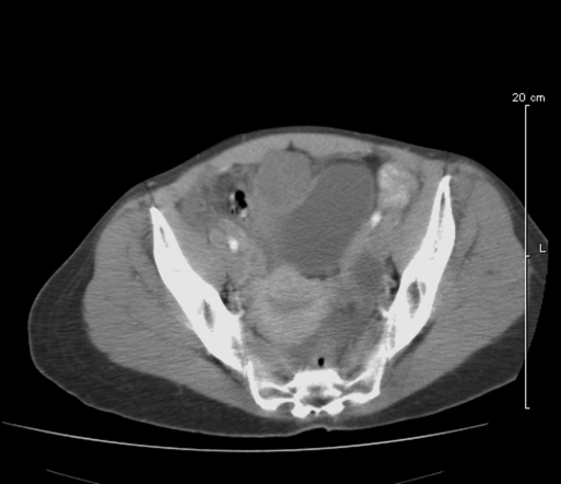
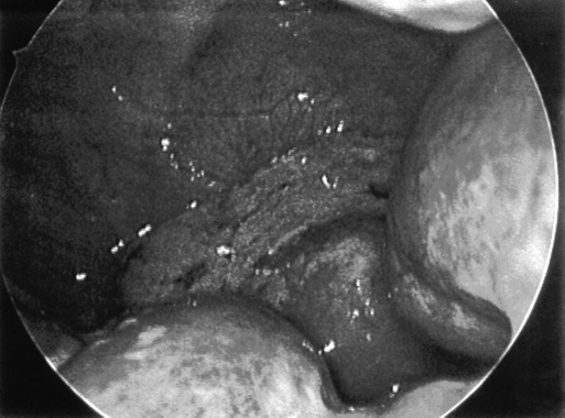
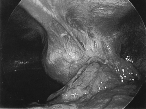
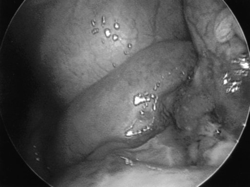
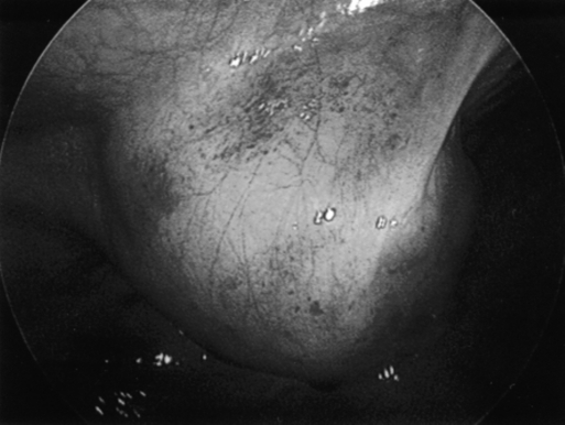
Methods: We present a case of a 51-year-old female presenting to our institution with complaints of abdominal pain for 36 hours. The patient was taken to the operating room for an acute appendicitis. Laparoscopy was performed, and gross purulence and appendiceal perforation were noted as well as a mass on the anterior abdominal wall. Based on the location of the mass, we converted to an open midline laparotomy to treat both the perforated appendicitis and to remove the mass.
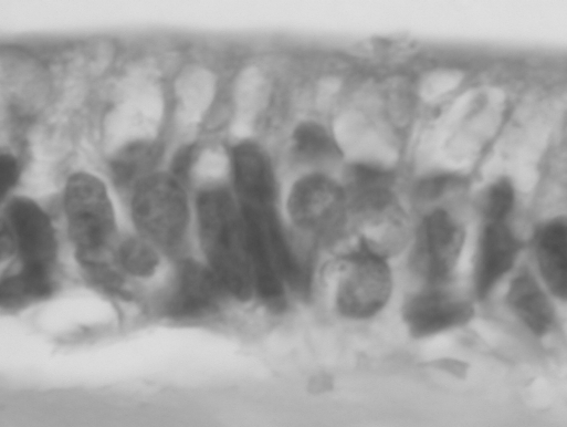
Results: Pathology confirmed the diagnosis of perforated appendicitis and a mucinous-producing urachal adenocarcinoma.
Discussion: Data support both open and laparoscopic approaches for appendicitis. This case, although rare, highlights the importance of laparoscopy in a complete and thorough examination of the abdominal cavity. A standard right lower quadrant incision for an open technique would likely have resulted in omission of this lesion, and the patient would have presented at a more typical late stage of her cancer development with significantly more morbidity.
Figures
Similar articles
-
Robotic assisted laparoscopic partial cystectomy and urachal resection for urachal adenocarcinoma.Int Braz J Urol. 2009 Sep-Oct;35(5):609. doi: 10.1590/s1677-55382009000500014. Int Braz J Urol. 2009. PMID: 19860941
-
[Chain of errors in laparoscopic appendectomy].Cir Cir. 2012 Jul-Aug;80(4):379-84. Cir Cir. 2012. PMID: 23374388 Spanish.
-
Laparoscopic management of symptomatic urachal remnants in adulthood.Asian J Surg. 2015 Apr;38(2):85-90. doi: 10.1016/j.asjsur.2014.04.009. Epub 2014 Jun 16. Asian J Surg. 2015. PMID: 24947766
-
[Adenocarcinomas of the urachus].Arch Esp Urol. 1991 Jan-Feb;44(1):31-6. Arch Esp Urol. 1991. PMID: 1648342 Review. Spanish.
-
Limited, local, extracolonic spread of mucinous appendiceal adenocarcinoma after perforation with formation of a malignant appendix-to-sigmoid fistula: Case report and literature review.World J Gastroenterol. 2016 Oct 14;22(38):8624-8630. doi: 10.3748/wjg.v22.i38.8624. World J Gastroenterol. 2016. PMID: 27784975 Free PMC article. Review.
Cited by
-
Urachal adenocarcinoma that metastasized to breast was misinterpreted as primary breast mucinous carcinoma: A rare case report and literature review.Medicine (Baltimore). 2016 Aug;95(35):e4612. doi: 10.1097/MD.0000000000004612. Medicine (Baltimore). 2016. PMID: 27583877 Free PMC article. Review.
-
Biomarkers in Urachal Cancer and Adenocarcinomas in the Bladder: A Comprehensive Review Supplemented by Own Data.Dis Markers. 2018 Mar 12;2018:7308168. doi: 10.1155/2018/7308168. eCollection 2018. Dis Markers. 2018. PMID: 29721106 Free PMC article. Review.
-
Surgical treatment of urachal adenocarcinoma with lung metastasis: A case report and literature review.Thorac Cancer. 2024 Dec;15(35):2509-2513. doi: 10.1111/1759-7714.15481. Epub 2024 Oct 27. Thorac Cancer. 2024. PMID: 39462217 Free PMC article. Review.
References
-
- Minevich E, Sheldon CA. In: Greenfield LJ, et al. ed. Surgery: Scientific Principles & Practice. 3rd ed. Philadelphia, PA: Lippincott Williams & Wilkins; 2001; 2047
-
- Ohira S, Shiohara S, Itoh K, Ashida T, Fukushima M, Konishi I. Urachal Adenocarcinoma metastatic to the ovaries: case report and literature review. Int J Gynelcol Pathol. 2003; 22 (2): 189–193 - PubMed
-
- Ichikawa T. Remote results of bladder tumors. Jpn J Urol. 1958; 49: 602–610
-
- Jacobo E, Loening S, Schmidt JD, et al. Primary adenocarcinoma of the bladder: a retrospective study of 20 patients. J Urol. 1977; 117: 54–56 - PubMed
-
- Thomas DG, Ward AM, Williams JL. A study of 52 cases of adenocarcinoma of the bladder. Br J Urol. 1971; 43: 4–15 - PubMed
Publication types
MeSH terms
LinkOut - more resources
Full Text Sources
Medical