Review
. 2007 Jan-Feb;52(1):97-105.
doi: 10.1016/j.survophthal.2006.10.006.

Ophthalmic manifestations and histopathology of infantile nephropathic cystinosis: report of a case and review of the literature

Affiliations
Review

Ophthalmic manifestations and histopathology of infantile nephropathic cystinosis: report of a case and review of the literature

Ekaterini Tsilou et al. Surv Ophthalmol. 2007 Jan-Feb.

Abstract

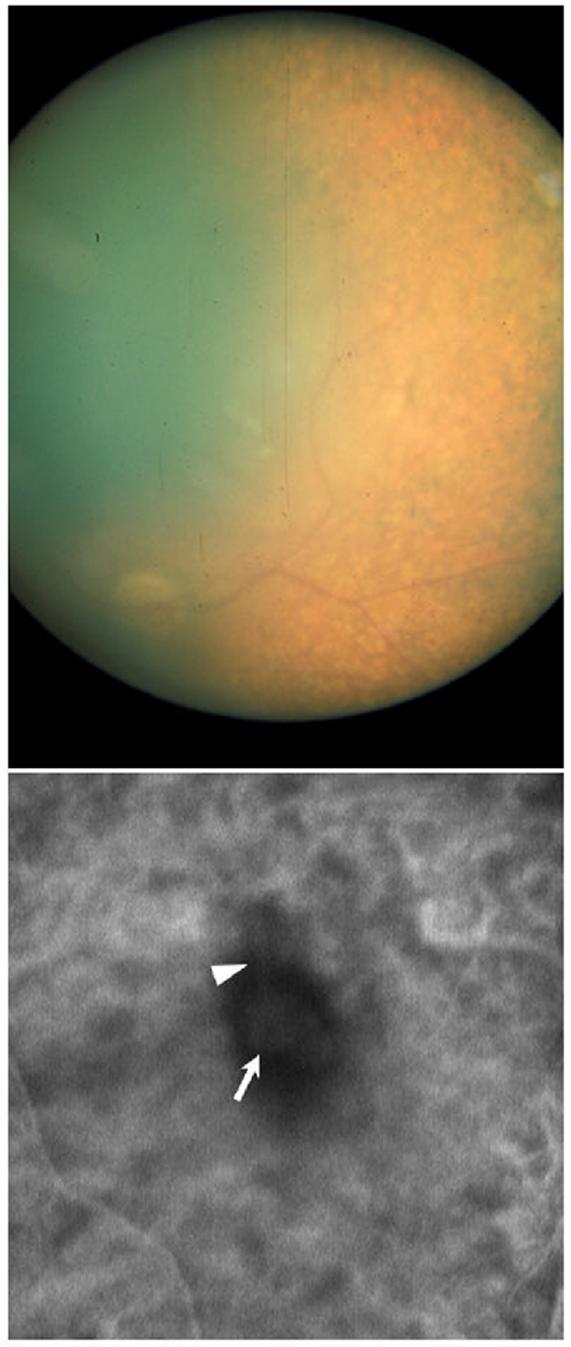
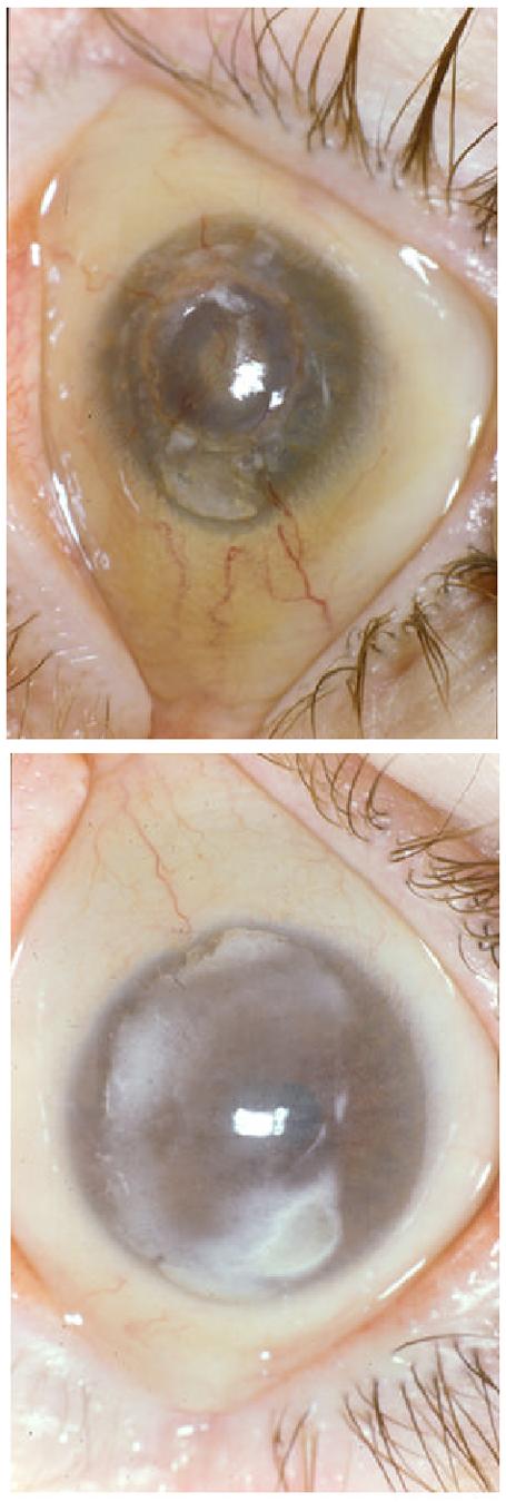
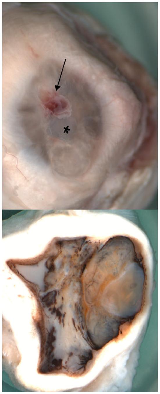
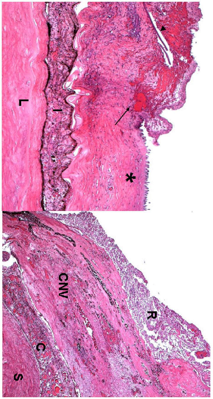
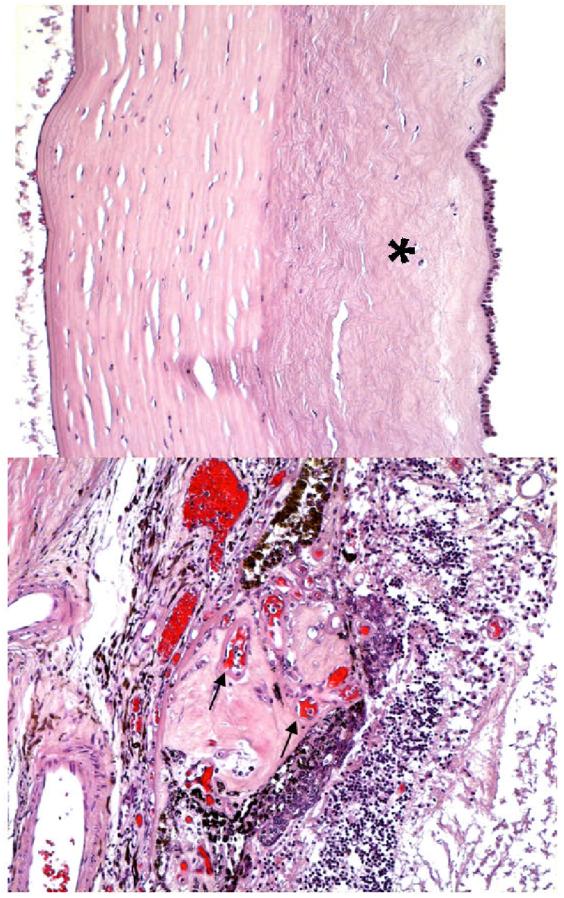
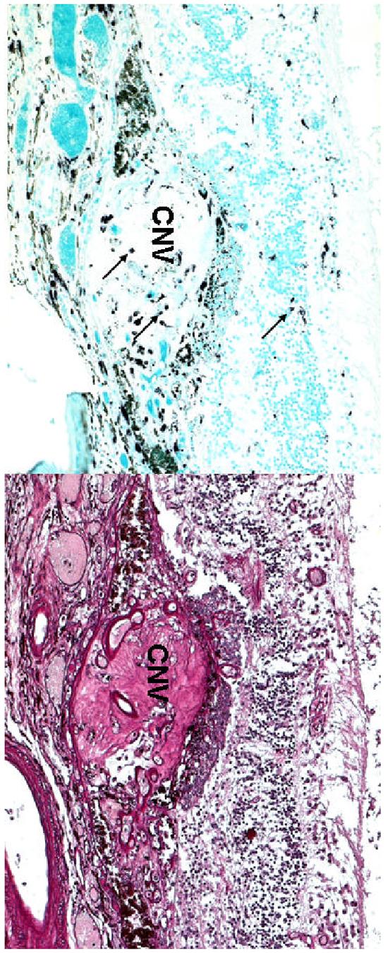
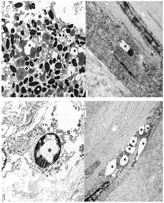
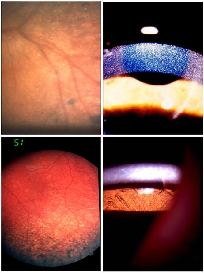
Cystinosis is a rare autosomal recessive metabolic disorder characterized by the intracellular accumulation of cystine, the disulfide of the amino acid cysteine, in many organs and tissues. Infantile nephropathic cystinosis is the most severe phenotype. Corneal crystal accumulation and pigmentary retinopathy were originally the most commonly described ophthalmic manifestations, but successful kidney transplantation significantly changed the natural history of the disease. As cystinosis patients now live longer, long-term complications in extrarenal tissues, including the eye, have become apparent. A case of an adult patient with infantile nephropathic cystinosis is reported. He presented with many long-term ocular complications of cystinosis. After 4 years of follow-up, the patient died from sepsis. Pathology of the phthisical eyes demonstrated numerous electron-transparent polygonal spaces, bounded by single membrane, in corneal cells, retinal pigment epithelial cells, and even choroidal endothelial cells. The ophthalmic manifestations and pathology of infantile nephropathic cystinosis are discussed and reviewed in light of the current report and other cases in the literature.


Figures

Figure 1
Figure 1
Fundus photograph of the right eye (A) and ICG-V image of the left fundus (B). Typical retinal pigmentary changes are obvious in the retinal periphery of the right eye (A). A subfoveal choroidal neovascular membrane (arrow) with contiguous feeding vessel (arrowhead) are evident in the left eye (B). Please note the poor quality of the fundus photograph, due to the cornea opacification and the inadequate dilation achieved secondary to the posterior synechiae.
Figure 2
Figure 2
Slit lamp photograph of right eye (A) and left eye (B). Calcification is present in both corneas. Severe cornea neovascularization is seen in the right cornea.
Figure 3
Figure 3
Macroscopic photographs of the right eye showing (A) a central corneal ulceration (asterisk) surrounded by a patch of hemorrhage (arrow); (B) a total obliterated anterior chamber, compressed ciliary body, hypermature cataract lens (L), vitreous cavity filled with white necrotic material, partial choroidal detachment, and thickened sclera (S).
Figure 4
Figure 4
Microphotographs of the right eye showing (A) the corneal ulcer (asterisk), surrounded by hemorrhages (arrow), neovascular fibrous tissue with linear calcified deposits (arrowhead), (I = iris, L= lens); (B) a thick layer of subretinal fibrosiscomposed of many small congested vessels (CNV) beneath the necrotic retina. (R = retina, CNV = choroidal neovascularization, C = choroid; S = sclera; hematoxylin-eosin, A x 50, B x 100)
Figure 5
Figure 5
Microphotographs of the left eye showing (A) a thin, delicate collagen scars (asterisk) replaced Bowman’s membrane and the anterior half of the stroma; (B) a large choroidal nodule composed of multiple small congested vessels (arrows) beneath the disrupted RPE and disorganized neuro-retina. (hematoxylin-eosin, A x 100,B x 200)
Figure 6
Figure 6
Microphotographs of the left eye showing (A) scattered macrophages (arrows) located in the choroidal neovascular nodule (CNV) and retina. (B) multiple small vascular lumens in the CNV. (A, abc immunohistochemistry, x 100; B, Periodic Acid Schiff, x 100)
Figure 7
Figure 7
Transmission electron micrographs showing (A) a prominent, polygonal electron-transparent area (asterisk) and adjacent lysosome bounded by the same single membrane in the cytoplasm of a keratocyte. (B) multiple polygonal electron-transparent structures (asterisks) in the cytoplasm of another keratocyte (N, nucleus). (C) prominent phagosomes containing polygonal electron-transparent structures (asterisks) bounded by a single membrane in the cytoplasm of a RPE cell; (D) intracytoplasmic electron-transparent area bounded by a single membrane (asterisk) in a choroidal fibroblast .
Figure 8
Figure 8
Ophthalmic manifestations of infantile nephropathic cystinosis: corneal crystals (A), iris crystals (B), retinal crystals (C), peripheral retinal pigmentary changes (D).

References

    1. ___ Feature photo. Cystinosis with extensive choroidal involvement. Arch Ophthalmol. 1965;74:868–9. - PubMed
    1. Aberhalden E. Familiare cystidiathesis. ZtschrPhysiol Chem. 1903;38:557.
    1. Almond PS, Matas AJ, Nakhleh RE, et al. Renal transplantation for infantile cystinosis: long-term follow-up. J Pediatr Surg. 1993;28:232–8. - PubMed
    1. Alsuhaibani AH, Khan AO, Wagoner MD. Confocal microscopy of the cornea in nephropathic cystinosis. Br J Ophthalmol. 2005;89:1530–1. - PMC - PubMed
    1. Anikster Y, Lucero C, Guo J, et al. Ocular nonnephropathic cystinosis: clinical, biochemical, and molecular correlations. Pediatr Res. 2000;47:17–23. - PubMed