

Angiography of primary central nervous system angiitis of childhood: conventional angiography versus magnetic resonance angiography at presentation
- PMID: 17213414
- PMCID: PMC8134080
Angiography of primary central nervous system angiitis of childhood: conventional angiography versus magnetic resonance angiography at presentation
Abstract
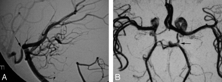
Background and purpose: To systematically analyze conventional angiographic (CA) features of children with primary central nervous system angiitis (cPACNS), to compare and correlate CA and MR angiography (MRA) lesion characteristics, and to define the sensitivity and specificity of MRA with CA as a reference standard.
Methods: A retrospective, single-center cohort study of consecutive patients with cPACNS was performed. Patients with CA and MRA studies at diagnosis were included. Imaging studies were blindly reviewed by 2 neuroradiologists using a standard analysis protocol. CA and MRA studies were compared using nonparametric analysis.
Results: Of 45 patients with MRA at diagnosis, there were 25 for whom CA and MRA studies were performed within 1 month of each other. These comprised the study group. The CA distribution of lesions was multifocal (76%) and proximal (86%) (P < .05) with a trend toward unilaterality (P = .06) with anterior circulation involvement (P = .08). The sensitivity and specificity of MRA for CA abnormality was 70% and 98%, respectively. There was no significant difference between MRA and CA for lesion detection or characterization (P = .87), and the modalities showed a fair correlation (kappa = 0.4).
Conclusion: Angiographic lesions are multifocal and occur proximally and unilaterally within the anterior circulation. There is no significant difference in the ability of MRA to detect and characterize lesions when compared with CA.
Figures


Similar articles
-
MR imaging and angiography of primary CNS vasculitis of childhood.AJNR Am J Neuroradiol. 2006 Jan;27(1):192-9. AJNR Am J Neuroradiol. 2006. PMID: 16418382 Free PMC article.
-
Investigation of childhood central nervous system vasculitis: magnetic resonance angiography versus catheter cerebral angiography.Dev Med Child Neurol. 2010 Sep;52(9):863-7. doi: 10.1111/j.1469-8749.2009.03591.x. Epub 2010 Jan 28. Dev Med Child Neurol. 2010. PMID: 20132140
-
Magnetic resonance angiography in childhood arterial brain infarcts: a comparative study with contrast angiography.Stroke. 2002 May;33(5):1280-5. doi: 10.1161/01.str.0000014504.18199.0d. Stroke. 2002. PMID: 11988604 Clinical Trial.
-
[Magnetic resonance angiography in inflammatory brain diseases].Radiologe. 2000 Nov;40(11):1077-89. doi: 10.1007/s001170050880. Radiologe. 2000. PMID: 11147323 Review. German.
-
Childhood central nervous system vasculitis.Handb Clin Neurol. 2013;112:1065-78. doi: 10.1016/B978-0-444-52910-7.00024-6. Handb Clin Neurol. 2013. PMID: 23622312 Review.
Cited by
-
Cerebral aneurysms in children: are we talking about a single pathological entity?Childs Nerv Syst. 2010 Oct;26(10):1329-35. doi: 10.1007/s00381-010-1205-z. Epub 2010 Jul 13. Childs Nerv Syst. 2010. PMID: 20625744
-
Primary angiitis of the central nervous system in a 7-month-old infant.Childs Nerv Syst. 2017 Feb;33(2):223-225. doi: 10.1007/s00381-017-3339-8. Epub 2017 Jan 12. Childs Nerv Syst. 2017. PMID: 28083640 No abstract available.
-
Strategies for treatment of childhood primary angiitis of the central nervous system.Neurol Neuroimmunol Neuroinflamm. 2019 May 3;6(4):e567. doi: 10.1212/NXI.0000000000000567. eCollection 2019 Jul. Neurol Neuroimmunol Neuroinflamm. 2019. PMID: 31355303 Free PMC article. Review.
-
Primary Angiitis of the Central Nervous System - Diagnosis and Management.Ann Indian Acad Neurol. 2022 Nov-Dec;25(6):1009-1018. doi: 10.4103/aian.aian_368_22. Epub 2022 Aug 10. Ann Indian Acad Neurol. 2022. PMID: 36911437 Free PMC article.
-
Imaging in childhood arterial ischaemic stroke.Neuroradiology. 2010 Jun;52(6):577-89. doi: 10.1007/s00234-010-0704-7. Epub 2010 May 6. Neuroradiology. 2010. PMID: 20445969 Review.
References
-
- Vollmer TL, Guarnaccia J, Harrington W, et al. Idiopathic granulomatous angiitis of the central nervous system; diagnostic challenges. Arch Neurol 1993;50:925–30 - PubMed
-
- Ganesan V, Savy L, Chong WK, et al. Conventional cerebral angiography in children with stroke. Paediatr Neurol 1999;20:38–42 - PubMed
-
- Husson B, Lasjaunias P. Radiological approach to disorders of arterial brain vessels associated with childhood arterial stroke—a comparison between MRA and contrast angiography. Pediatr Radiol 2004;34:10–15 - PubMed
Publication types
MeSH terms
LinkOut - more resources
Full Text Sources