

Brain AVM embolization with Onyx
- PMID: 17213451
- PMCID: PMC8134108
Brain AVM embolization with Onyx
Abstract
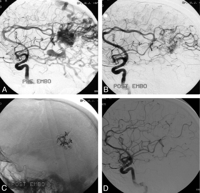
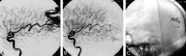
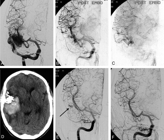
Background and purpose: To report the initial experience by using a new liquid embolic agent (Onyx) for embolization of brain arteriovenous malformations (AVMs).
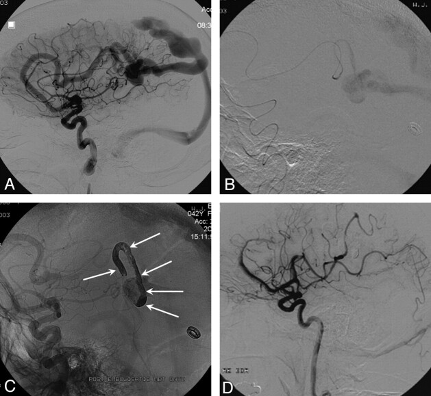
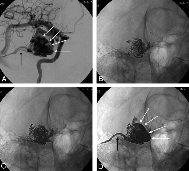
Methods: Between May 2000 and December 2005, 44 patients with brain AVMs were embolized with Onyx. There were 18 women and 26 men with a mean age of 42.4 years (median 44, range 14-71 years). Clinical presentation included seizures in 26 patients (59%), hemorrhage from the AVM in 13 patients (30%), subarachnoid hemorrhage from a concomitant aneurysm in 3 patients (7%), visual disturbances in 1 patient (2.3%), and in 1 patient (2.3%) the AVM was an incidental finding. Mean estimated size of the AVM was 3.9 cm (median 4, range 2-7 cm).
Results: In 44 patients, 52 embolization procedures were performed with 138 feeding pedicles embolized, ranging from 1 to 7 per patient. Average estimated size reduction was 75% (median 80%, range 40%-100%). Total obliteration was achieved in 7 AVMs (16%), and partial embolization was followed by surgery in 10 patients and by radiosurgery in 20 patients. Complications occurred in 6 patients, leading to death in 1 patient (mortality 2.3%) and to permanent disability in 2 patients (morbidity 4.6%).
Conclusion: Onyx is feasible and safe in the embolization of brain AVMs. Complete obliteration can be achieved in small AVMs. Large AVMs can be adequately reduced in size for additional surgical or radiosurgical treatment.
Figures
References
-
- Richling B, Killer M. Endovascular management of patients with cerebral arteriovenous malformations. Neurosurg Clin N Am 2000;11:123–45 - PubMed
-
- Henkes H, Nahser HC, Berg-Dammer E, et al. Endovascular therapy of brain AVMs prior to radiosurgery. Neurol Res 1998;20:479–92 - PubMed
-
- Gobin YP, Laurent A, Merienne L, et al. Treatment of brain arteriovenous malformations by embolization and radiosurgery. J Neurosurg 1996;85:19–28 - PubMed
-
- Spetzler RF, Martin NA, Carter LP, et al. Surgical management of large AVM’s by staged embolization and operative excision. J Neurosurg 1987;67:17–28 - PubMed
-
- Jahan R, Murayama Y, Gobin YP, et al. Embolization of arteriovenous malformations with Onyx: clinicopathological experience in 23 patients. Neurosurgery 2001;48:984–95 - PubMed
Publication types
MeSH terms
Substances
LinkOut - more resources
Full Text Sources