. 2007 Apr;81(7):3087-96.
doi: 10.1128/JVI.02032-06. Epub 2007 Jan 10.

Classical swine fever virus Npro interacts with interferon regulatory factor 3 and induces its proteasomal degradation

Affiliations

Classical swine fever virus Npro interacts with interferon regulatory factor 3 and induces its proteasomal degradation

Oliver Bauhofer et al. J Virol. 2007 Apr.

Abstract

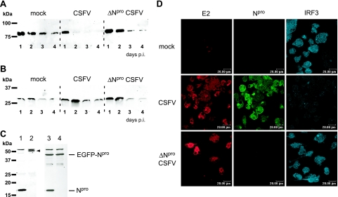
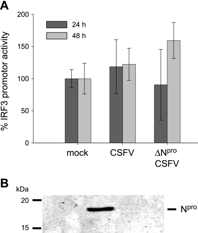
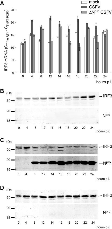
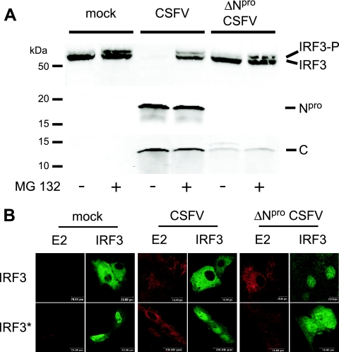
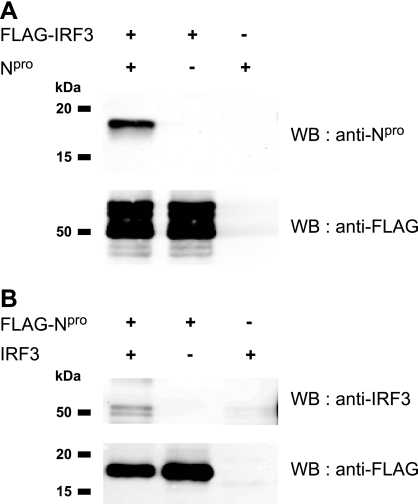
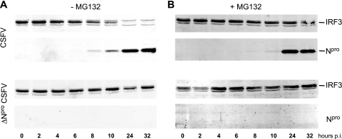
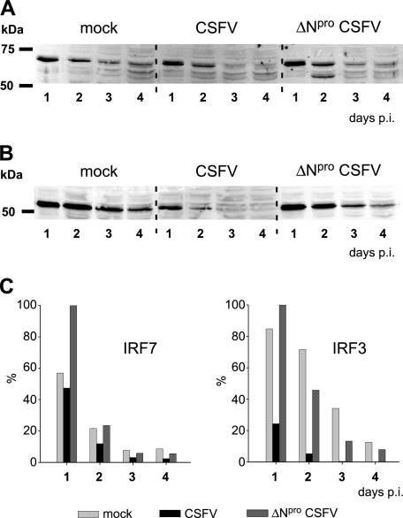
Viruses have evolved a multitude of strategies to subvert the innate immune system by interfering with components of the alpha/beta interferon (IFN-alpha/beta) induction and signaling pathway. It is well established that the pestiviruses prevent IFN-alpha/beta induction in their primary target cells, such as epitheloidal and endothelial cells, macrophages, and conventional dendritic cells, a phenotype mediated by the viral protein N(pro). Central players in the IFN-alpha/beta induction cascade are interferon regulatory factor 3 (IRF3) and IRF7. Recently, it was proposed that classical swine fever virus (CSFV), the porcine pestivirus, induced the loss of IRF3 by inhibiting the transcription of IRF3 mRNA. In the present study, we show that endogenous IRF3 and IRF3 expressed from a cytomegalovirus (CMV) promoter are depleted in the presence of CSFV by means of N(pro), while CSFV does not inhibit CMV promoter-driven protein expression. We also demonstrate that CSFV does not reduce the transcriptional activity of the IRF3 promoter and does not affect the stability of IRF3 mRNA. In fact, CSFV N(pro) induces proteasomal degradation of IRF3, as demonstrated by proteasome inhibition studies. Furthermore, N(pro) coprecipitates with IRF3, suggesting that the proteasomal degradation of IRF3 is induced by a direct or indirect interaction with N(pro). Finally, we show that N(pro) does not downregulate IRF7 expression.


Figures

FIG. 1.
FIG. 1.
CSFV infection induces loss of CMV-driven IRF3, a mechanism dependent on the presence of Npro. PK-15 cells were transfected with pEGFP-IRF3 (A) or with pEGFP-N1 (B). Twenty-four hours after transfection, the cells were mock treated or infected with CSFV or ΔNpro CSFV at an MOI of 0.2 TCID50/cell. On the indicated days p.i., the cells were lysed and extracts were analyzed for EGFP-IRF3 (A) and EGFP (B) expression by Western blotting with the anti-EGFP MAb JL-8. (C) IRF3 expression was analyzed in the clonal PK15-EGFP (lanes 1 and 2) and PK15-EGFP-Npro (lanes 3 and 4) cell lines. Cells were infected with CSFV at an MOI of 2 TCID50/cell (lanes 1 and 3) or mock treated (lanes 2 and 4). Twenty-four hours p.i., cell extracts were analyzed for IRF3, EGFP-Npro, and Npro expression by Western blotting using the rabbit anti-IRF3 and anti-Npro sera, respectively. The IRF3 protein in the PK15-EGFP extract (lane 2) is indicated with an arrowhead. (D) Endogenous IRF3 was analyzed in DC after mock, CSFV, and ΔNpro CSFV infection. After 4 days of differentiation, DC were transferred into chamber slides and mock infected or infected with CSFV or ΔNpro CSFV at an MOI of 20 TCID50/cell (based on the virus titer on SK-6 cells). Confocal microscopy was performed 48 h p.i. For this purpose, the cells were fixed and stained for the viral proteins E2 and Npro with MAb HC/TC26 and with the rabbit anti-Npro serum, respectively, and for IRF3 with the rabbit anti-IRF3 serum as indicated.
FIG. 2.
FIG. 2.
Npro does not inhibit transcription of IRF3. (A) PK-15 cells were transfected with a plasmid encoding firefly luciferase under the control of the human IRF3 promoter and a second plasmid expressing Renilla luciferase under the SV40 promoter. Twenty-four hours after transfection, the cells were mock treated or infected with CSFV or ΔNpro CSFV. Cells were lysed 24 and 48 h p.i. and analyzed for the relative firefly and Renilla luciferase activities using the dual-luciferase assay system. The normalized relative induction is represented as a percentage of the promoter activity of mock-infected cells (mock = 100%). The error bars represent the standard deviations. (B) In parallel, protein extracts were analyzed for the presence of Npro by Western blotting using the rabbit anti-Npro serum.
FIG. 3.
FIG. 3.
Npro does not destabilize IRF3 mRNA. (A) The IRF3 mRNA contents of PK-15 cells at different times after mock treatment or infection with either CSFV or ΔNpro CSFV were measured by real-time RT-PCR. The amount of IRF3 mRNA is shown as CT (no RT)CT (RT-PCR), with error bars representing the 95% confidence interval. In parallel with the RNA extraction, cells were lysed at the indicated times, and the IRF3 and Npro proteins were analyzed by Western blotting in mock- (B), CSFV- (C), and ΔNpro CSFV-infected (D) cells using the rabbit anti-IRF3 and anti-Npro sera.
FIG. 4.
FIG. 4.
Npro does not prevent IRF3 phosphorylation or nuclear translocation of a constitutively active form of IRF3. (A) PK-15 cells were treated with 0.2 μM of the proteasome inhibitor MG132 (+) or left untreated (−) and infected with CSFV or ΔNpro CSFV at an MOI of 0.2 TCID50/cell or mock treated as indicated. Sixteen hours p.i., the cells were lysed and analyzed by Western blotting for endogenous IRF3 using MAb 34/1 and for the viral proteins Npro and capsid C using the respective rabbit antisera as described in Materials and Methods. (B) PK-15 cells were mock treated or infected with CSFV or ΔNpro CSFV at an MOI of 2 TCID50/cell and transfected with pFLAG-IRF3 (top row) or pFLAG-IRF3-S394,396D-ΔNES (IRF3*; bottom row). Sixteen hours after transfection, the cells were fixed and stained for the viral E2 protein and for the FLAG tag of IRF3 as indicated and analyzed by confocal microscopy.
FIG. 5.
FIG. 5.
IRF3 and Npro interact with each other. HEK 293T cells were transfected with plasmids pFLAG-IRF3 and pEAK-Npro for the expression of FLAG-tagged IRF3 and untagged Npro (A) or with pFLAG-Npro and pIRF3 for the expression of FLAG-tagged Npro and untagged IRF3 (B), either individually or mixed at a 1:1 ratio. The presence and absence of the expression plasmid are indicated by + and −, respectively. Twenty-four hours after transfection, the cells were lysed and the proteins were immunoprecipitated with anti-FLAG M2 agarose. The precipitated proteins were eluted from the agarose, separated by SDS-PAGE, and analyzed by Western blotting (WB) with rabbit anti-Npro serum, anti-IRF3 MAb 34/1, and anti-FLAG MAb as indicated.
FIG. 6.
FIG. 6.
Npro induces proteasomal degradation of IRF3. PK-15 cells were infected with CSFV (top) or with ΔNpro CSFV (bottom) at an MOI of 0.2 TCID50/cell in the absence (A) or in the presence (B) of 0.2 μM proteasome inhibitor MG132. At the indicated times p.i., the cells were lysed and the Npro and IRF3 contents were analyzed by Western blotting using the rabbit anti-Npro and anti-IRF3 sera, respectively.
FIG. 7.
FIG. 7.
CSFV Npro does not downregulate IRF7 expression. PK-15 cells were transfected with plasmid pFLAG-IRF7 (A) or pFLAG-IRF3 (B) for CMV promoter-driven expression of FLAG-tagged IRF7 or FLAG-tagged IRF3, respectively. Twenty-four hours after transfection, the cells were mock treated or infected with CSFV or ΔNpro CSFV at an MOI of 2 TCID50/cell. On the indicated days p.i., cells were lysed and extracts were analyzed for IRF7 (A) and IRF3 (B) expression by Western blotting using the rabbit anti-FLAG MAb. (C) The IRF7- and IRF3-specific signals from the Western blots shown in panels A and B were quantified with the Odyssey Imaging system and expressed as percentages of the strongest signal.

References

    1. Andrejeva, J., K. S. Childs, D. F. Young, T. S. Carlos, N. Stock, S. Goodbourn, and R. E. Randall. 2004. The V proteins of paramyxoviruses bind the IFN-inducible RNA helicase, mda-5, and inhibit its activation of the IFN-beta promoter. Proc. Natl. Acad. Sci. USA 101:17264-17269. - PMC - PubMed
    1. Andrejeva, J., E. Poole, D. F. Young, S. Goodbourn, and R. E. Randall. 2002. The p127 subunit (DDB1) of the UV-DNA damage repair binding protein is essential for the targeted degradation of STAT1 by the V protein of the paramyxovirus simian virus 5. J. Virol. 76:11379-11386. - PMC - PubMed
    1. Baigent, S. J., S. Goodbourn, and J. W. McCauley. 2004. Differential activation of interferon regulatory factors-3 and -7 by non-cytopathogenic and cytopathogenic bovine viral diarrhoea virus. Vet. Immunol. Immunopathol. 100:135-144. - PubMed
    1. Baigent, S. J., G. Zhang, M. D. Fray, H. Flick-Smith, S. Goodbourn, and J. W. McCauley. 2002. Inhibition of beta interferon transcription by noncytopathogenic bovine viral diarrhea virus is through an interferon regulatory factor 3-dependent mechanism. J. Virol. 76:8979-8988. - PMC - PubMed
    1. Balmelli, C., I. E. Vincent, H. Rau, L. Guzylack-Piriou, K. McCullough, and A. Summerfield. 2005. FcγRII-dependent sensitisation of natural interferon-producing cells for viral infection and interferon-alpha responses. Eur. J. Immunol. 35:2406-2415. - PubMed

Publication types

MeSH terms

Associated data