Electron tomography of nascent herpes simplex virus virions
- PMID: 17215293
- PMCID: PMC1865967
- DOI: 10.1128/JVI.02571-06
Electron tomography of nascent herpes simplex virus virions
Abstract
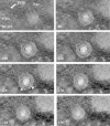
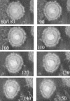
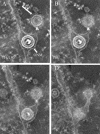
Cells infected with herpes simplex virus type 1 (HSV-1) were conventionally embedded or freeze substituted after high-pressure freezing and stained with uranyl acetate. Electron tomograms of capsids attached to or undergoing envelopment at the inner nuclear membrane (INM), capsids within cytoplasmic vesicles near the nuclear membrane, and extracellular virions revealed the following phenomena. (i) Nucleocapsids undergoing envelopment at the INM, or B capsids abutting the INM, were connected to thickened patches of the INM by fibers 8 to 19 nm in length and < or =5 nm in width. The fibers contacted both fivefold symmetrical vertices (pentons) and sixfold symmetrical faces (hexons) of the nucleocapsid, although relative to the respective frequencies of these subunits in the capsid, fibers engaged pentons more frequently than hexons. (ii) Fibers of similar dimensions bridged the virion envelope and surface of the nucleocapsid in perinuclear virions. (iii) The tegument of perinuclear virions was considerably less dense than that of extracellular virions; connecting fibers were observed in the former case but not in the latter. (iv) The prominent external spikes emanating from the envelope of extracellular virions were absent from perinuclear virions. (v) The virion envelope of perinuclear virions appeared denser and thicker than that of extracellular virions. (vi) Vesicles near, but apparently distinct from, the nuclear membrane in single sections were derived from extensions of the perinuclear space as seen in the electron tomograms. These observations suggest very different mechanisms of tegumentation and envelopment in extracellular compared with perinuclear virions and are consistent with application of the final tegument to unenveloped nucleocapsids in a compartment(s) distinct from the perinuclear space.
Figures


Similar articles
-
Identification of the Capsid Binding Site in the Herpes Simplex Virus 1 Nuclear Egress Complex and Its Role in Viral Primary Envelopment and Replication.J Virol. 2019 Oct 15;93(21):e01290-19. doi: 10.1128/JVI.01290-19. Print 2019 Nov 1. J Virol. 2019. PMID: 31391274 Free PMC article.
-
UL20 protein functions precede and are required for the UL11 functions of herpes simplex virus type 1 cytoplasmic virion envelopment.J Virol. 2007 Apr;81(7):3097-108. doi: 10.1128/JVI.02201-06. Epub 2007 Jan 10. J Virol. 2007. PMID: 17215291 Free PMC article.
-
Herpes simplex virus nucleocapsids mature to progeny virions by an envelopment --> deenvelopment --> reenvelopment pathway.J Virol. 2001 Jun;75(12):5697-702. doi: 10.1128/JVI.75.12.5697-5702.2001. J Virol. 2001. PMID: 11356979 Free PMC article.
-
Virus Assembly and Egress of HSV.Adv Exp Med Biol. 2018;1045:23-44. doi: 10.1007/978-981-10-7230-7_2. Adv Exp Med Biol. 2018. PMID: 29896661 Review.
-
Host Cell Signatures of the Envelopment Site within Beta-Herpes Virions.Int J Mol Sci. 2022 Sep 1;23(17):9994. doi: 10.3390/ijms23179994. Int J Mol Sci. 2022. PMID: 36077391 Free PMC article. Review.
Cited by
-
The Herpes Simplex Virus Protein pUL31 Escorts Nucleocapsids to Sites of Nuclear Egress, a Process Coordinated by Its N-Terminal Domain.PLoS Pathog. 2015 Jun 17;11(6):e1004957. doi: 10.1371/journal.ppat.1004957. eCollection 2015 Jun. PLoS Pathog. 2015. PMID: 26083367 Free PMC article.
-
Viral Infection at High Magnification: 3D Electron Microscopy Methods to Analyze the Architecture of Infected Cells.Viruses. 2015 Dec 3;7(12):6316-45. doi: 10.3390/v7122940. Viruses. 2015. PMID: 26633469 Free PMC article. Review.
-
Identification of functional domains within the essential large tegument protein pUL36 of pseudorabies virus.J Virol. 2007 Dec;81(24):13403-11. doi: 10.1128/JVI.01643-07. Epub 2007 Oct 10. J Virol. 2007. PMID: 17928337 Free PMC article.
-
Herpesviruses remodel host membranes for virus egress.Nat Rev Microbiol. 2011 May;9(5):382-94. doi: 10.1038/nrmicro2559. Nat Rev Microbiol. 2011. PMID: 21494278 Review.
-
The capsid protein encoded by U(L)17 of herpes simplex virus 1 interacts with tegument protein VP13/14.J Virol. 2010 Aug;84(15):7642-50. doi: 10.1128/JVI.00277-10. Epub 2010 May 26. J Virol. 2010. PMID: 20504930 Free PMC article.
References
Publication types
MeSH terms
Grants and funding
LinkOut - more resources
Full Text Sources
Other Literature Sources
Medical

