. 2007 Jan 16;104(3):991-6.
doi: 10.1073/pnas.0610270104. Epub 2007 Jan 10.

Mitochondrial dysfunction in the pathogenesis of Ullrich congenital muscular dystrophy and prospective therapy with cyclosporins

Affiliations

Mitochondrial dysfunction in the pathogenesis of Ullrich congenital muscular dystrophy and prospective therapy with cyclosporins

Alessia Angelin et al. Proc Natl Acad Sci U S A. .

Abstract

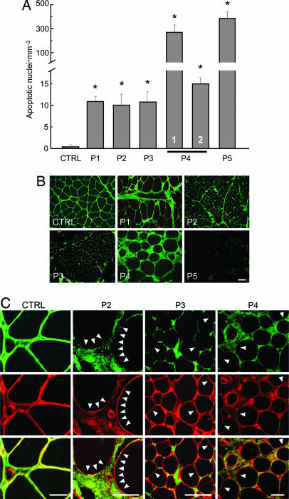
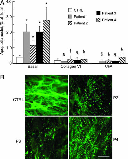
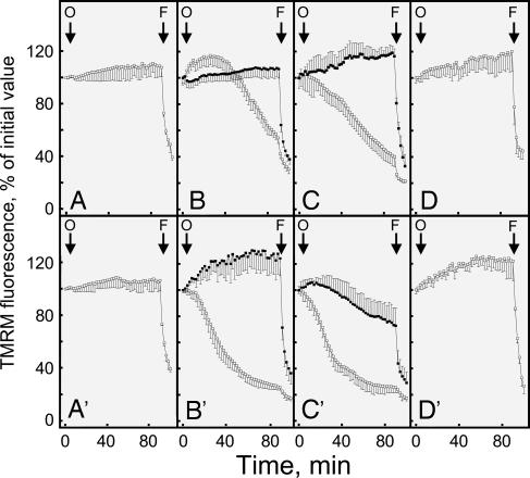
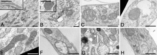
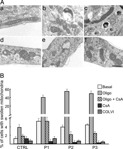
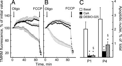
Ullrich congenital muscular dystrophy is a severe genetically and clinically heterogeneous muscle disorder linked to collagen VI deficiency. The pathogenesis of the disease is unknown. To assess the potential role of mitochondrial dysfunction in the onset of muscle fiber death in this form of dystrophy, we studied biopsies and myoblast cultures obtained from patients with different genetic defects of collagen VI and variable clinical presentations of the disease. We identified a latent mitochondrial dysfunction in myoblasts from patients with Ullrich congenital muscular dystrophy that matched an increased occurrence of spontaneous apoptosis. Unlike those in myoblasts from healthy donors, mitochondria in cells from patients depolarized upon addition of oligomycin and displayed ultrastructural alterations that were worsened by treatment with oligomycin. The increased apoptosis, the ultrastructural defects, and the anomalous response to oligomycin could be normalized by Ca(2+) chelators, by plating cells on collagen VI, and by treatment with cyclosporin A or with the specific cyclophilin inhibitor methylAla(3)ethylVal(4)-cyclosporin, which does not affect calcineurin activity. Here we demonstrate that mitochondrial dysfunction plays an important role in muscle cell wasting in Ullrich congenital muscular dystrophy. This study represents an essential step toward a pharmacological therapy of Ullrich congenital muscular dystrophy with cyclosporin A and methylAla(3)ethylVal(4) cyclosporin.


Conflict of interest statement

The authors declare no conflict of interest.

Figures

Fig. 1.
Fig. 1.
Incidence of apoptosis and expression of collagen VI in muscle biopsies from a healthy donor and UCMD patients. (A) Biopsies from a healthy donor (CTRL) and five UCMD patients (P1–P5) were scored for the presence of apoptotic nuclei with the TUNEL reaction. For patient 4, columns 1 and 2 refer to biopsies taken at 4 and 9 years of age, respectively. ∗, P < 0.01 for each patient vs. healthy donor. (B) Muscle biopsies from a healthy donor (CTRL) and five UCMD patients (P1–P5) were studied by immunofluorescence for collagen VI. (Scale bar, 20 μm.) (C) Muscle biopsies from a healthy donor (CTRL) and three UCMD patients (P2–P4) were studied by immunofluorescence for collagen VI (COLVI, green fluorescence) (Top) and perlecan (red fluorescence) (Middle), and a merge is shown in Bottom. Arrowheads mark examples of a lack of collagen VI staining at the basal lamina. (Scale bars, 20 μm.)
Fig. 2.
Fig. 2.
Incidence of apoptosis and expression of collagen VI in muscle cell cultures from a healthy donor and UCMD patients. (A) Primary myoblast cultures from a healthy donor (CTRL) and four UCMD patients were plated on plastic dishes or on collagen VI and scored for the presence of apoptotic nuclei by the TUNEL reaction. Where indicated, cells plated on plastic dishes were incubated with 1.6 μM CsA for 2 h. Data are the mean of three independent experiments ± SD. *, P < 0.01 for each patient vs. healthy donor; §, not significant for each patient vs. healthy donor. (B) Collagen VI was detected by immunofluorescence in cultured myoblasts from a healthy donor (CTRL) and three UCMD patients (P2–P4). Note that in the healthy donor collagen VI is secreted and organized in a dense fibrillar network, whereas in the UCMD patients collagen VI staining is severely reduced, with residual labeling in the form of small dots. (Scale bar, 20 μm.)
Fig. 3.
Fig. 3.
Changes of mitochondrial TMRM fluorescence induced by oligomycin in muscle cell cultures from a healthy donor and UCMD patients. Myoblasts from two healthy donors (A and A′) and from UCMD patients 1 (B–D) and 2 (B′–D′) were loaded with TMRM and studied as described in Materials and Methods. Cells were seeded on glass (A–C and A′–C′) or on collagen VI (D and D′). Where indicated by arrows, 6 μM oligomycin (O) and 4 μM carbonyl cyanide p-trifluoromethoxyphenylhydrazone (FCCP) (F) were added in the absence of further treatments (open symbols) or after treatment for 30 min with 1.6 μM CsA or 5 μM 1,2-bis(2-aminophenoxy)ethane-N,N,N′,N′-tetraacetic acid tetraacetoxymethyl ester (filled symbols in B and B′ and C and C′, respectively). Similar results were obtained with cells from patients 3 and 4, and the results shown are representative of at least five repeats per patient.
Fig. 4.
Fig. 4.
Mitochondrial ultrastructure in muscle cell cultures from healthy donors and UCMD patients. (A–E) Electron micrographs of myoblasts from a healthy donor (A) and UCMD patients 1, 2, and 3 (B, C, and D, respectively) plated on plastic or on collagen VI (patient 2, E). (A and B Insets) Higher magnifications from the same samples. (F–H) Representative examples of swollen mitochondria found in the myoblasts of patients 1 (F), 2 (G), and 3 (H). For further details and for a morphometric analysis see Results. (Scale bars, 300 nm.)
Fig. 5.
Fig. 5.
Effects of oligomycin, CsA, and collagen VI on mitochondrial ultrastructure in healthy donor and UCMD patient cell cultures. (A) Electron micrographs of myoblasts from a healthy donor (a–c) and UCMD patient 2 (d–f) plated on plastic in the absence (a and d) or presence of 6 μM oligomycin (b and e) or 6 μM oligomycin plus 1.6 μM CsA (c and f). (Scale bar, 400 nm.) (B) Percentage of cells with swollen mitochondria in the myoblasts of a healthy donor (CTRL) or UCMD patients 1, 2, and 3 (P1–P3) plated on plastic in the absence (Basal) or presence of 6 μM oligomycin (Oligo), 1.6 μM CsA, or both or of the same cells plated on collagen VI (COLVI). Data are the mean of three independent experiments ± SD.
Fig. 6.
Fig. 6.
Effects of CsA and Debio 025 on mitochondrial TMRM fluorescence and incidence of apoptosis in myoblast cultures from UCMD patients. (A and B) Myoblasts from UCMD patients 1 (A) and 4 (B) were loaded with TMRM and studied as described in Materials and Methods. Cells were seeded on glass, and, where indicated by arrows, 6 μM oligomycin (Oligo) and 4 μM carbonyl cyanide p-trifluoromethoxyphenylhydrazone (FCCP) were added in the absence of further treatments (open symbols) or after treatment for 30 min with 1.6 μM CsA (filled squares) or 1.6 μM Debio 025 (filled triangles). (C) Myoblasts from patients 1 (P1) and 4 (P4) were cultured on plastic dishes and scored for the presence of TUNEL-positive nuclei either in the absence of further treatments (Basal) or after incubation for 2 h with 1.6 μM CsA or 1.6 μM Debio 025. Data are the mean of at least four independent experiments ± SD. *, P < 0.01 compared with basal condition; §, not significant for Debio 025 vs. CsA.

References

    1. Lampe AK, Bushby KM. J Med Genet. 2005;42:673–685. - PMC - PubMed
    1. Bethlem J, Wijngaarden GK. Brain. 1976;99:91–100. - PubMed
    1. Merlini L, Morandi L, Granata C, Ballestrazzi A. Neuromuscul Disord. 1994;4:503–511. - PubMed
    1. Pepe G, Giusti B, Bertini E, Brunelli T, Saitta B, Comeglio P, Bolognese A, Merlini L, Federici G, Abbate R, et al. Biochem Biophys Res Commun. 1999;258:802–807. - PubMed
    1. de Visser M, de Voogt WG, la Riviere GV. Muscle Nerve. 1992;15:591–596. - PubMed

Publication types