Comparative Study
. 2007 Jan 10;27(2):431-45.
doi: 10.1523/JNEUROSCI.4979-06.2007.

Hepatocyte growth factor acts as a motogen and guidance signal for gonadotropin hormone-releasing hormone-1 neuronal migration

Affiliations
Comparative Study

Hepatocyte growth factor acts as a motogen and guidance signal for gonadotropin hormone-releasing hormone-1 neuronal migration

Paolo Giacobini et al. J Neurosci. .

Abstract

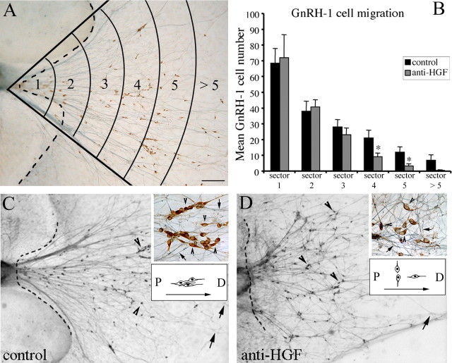
Reproduction in mammals is under the control of the hypothalamic neuropeptide gonadotropin hormone-releasing hormone-1 (GnRH-1). GnRH-1-secreting neurons originate during embryonic development in the nasal placode and migrate into the forebrain along olfactory nerves. Gradients of secreted molecules may play a role in this migratory process. In this context, hepatocyte growth factor (HGF) is a potential candidate, because it promotes cell motility in developing brain and has been shown previously to act as a motogen on immortalized GnRH-1 neurons (GN11). In this study, the role of HGF and its receptor Met during development of the GnRH-1 system was examined. GnRH-1 cells express Met during their migration and downregulate its expression once they complete this process. Tissue-type plasminogen activator (tPA), a known HGF activator, is also detected in migratory GnRH-1 neurons. Consistent with in vivo expression, HGF is present in nasal explants, and GnRH-1 neurons express Met. HGF-neutralizing antibody was applied to explants to examine the role of the endogenous growth factor. Migration of GnRH-1 cells and olfactory axon outgrowth were significantly reduced, in line with disruption of a guidance gradient. Exogenous application of HGF to explants increased the distance that GnRH-1 cells migrated, suggesting that HGF also acts as a motogen to GnRH-1 neurons. Functional experiments, performed on organotypic slice cultures, show that creation of an opposing HGF gradient inhibits GnRH-1 neuronal migration. Finally, tPA(-/-):uPA(-/-) (urokinase-type plasminogen activator(-/-)) knock-out mice exhibit strong reduction of the GnRH-1 cell population. Together, these data indicate that HGF signaling via Met receptor influences the development of GnRH-1.


Figures

Figure 1.
Figure 1.
Nasal regions express HGF and its receptor Met during embryonic development. Schematic of an E11.5–E12.5 head [fb, OE, presumptive VNO, tongue (t), and third ventricle (III) are depicted]. The dashed line indicates the boundary between nose and brain and represents the region taken for nasal RNA isolation in B. B, Gel documentation of products produced by RT-PCR amplification using specific primers for c-met and HGF. Total RNA was isolated from nose and whole head at E11.5. Adult brain was used as positive control tissue. Transcripts for both c-met and HGF were detected in all samples but water. Western blot analysis showed that HGF is expressed in its active form in protein extracts of E12 noses as well in whole heads of E16, used as positive control tissue. Western blot was run under reducing conditions. Met expression was also detected in the same samples. C, D, Photographs of E14.5 sagittal sections after LCM. Representative pictures show examples of microdissected OE (C) and VNO (D). No other tissue was removed from the nasal section, and the remaining tissue was intact after the capture procedure. E, Total RNA isolated from dissected regions was subjected to RT-PCR. A fragment of the expected size (519 bp) was detected for c-met in the OE and in VNO. Expression of the olfactory marker EBF-2 (165 bp) confirmed the morphology of the dissected tissue. PCR using HGF primers showed the expected amplicon (314 bp) in the positive control lane (CNTR; E17.5 whole-embryo extracts) but not in OE or VNO. No PCR product was observed in reactions that omitted either reverse transcriptase or starting material (water). F, G, Sagittal section of an E14.5 mouse nose double stained for NCAM (expressed by olfactory/vomeronasal axons; F) and Met (G). Met immunoreactivity is distributed in the developing OE and VNO structures and along the olfactory/vomeronasal fibers. G,Inset, Colocalization between the two antigens. MW, Molecular weight; OB, olfactory bulb. Scale bars: (in C) C, D, 30 μm; (in F) F, G, 100 μm; G,inset, 22 μm. Asterisks indicate laser-captured areas.
Figure 2.
Figure 2.
Met receptor expression in GnRH-1 neurons correlates with migration. A–C, Sagittal sections of E12.5 mouse immunostained with indicated antibodies. A, Met-immunoreactive cells emerged from the developing VNO and migrated through the olfactory mesenchyme (arrows) toward the forebrain. Met staining is also evident along the vomeronasal fibers coming out of the VNO. Arrowheads indicate Met-ir vomeronasal fibers. B, C, Double-label immunofluorescence for Met (red; B) and GnRH-1 (green; C) indicates that migrating GnRH-1 neurons spanning across the nasal regions coexpress Met (C, inset, arrows). D–F, Coronal section of hypothalamic area of a PN10 mouse double labeled for GnRH-1 (red; arrows) and Met (green; arrowheads). E, F, High-power confocal analysis showed that GnRH-1-immunoreactive cells and fibers do not colocalize with Met-immunopositive elements at this stage. III, Third ventricle. Scale bars: A, 30 μm; (in B) B, C, 10 μm; (in B) D, 15 μm; C, inset, 10 μm; (in E) E, F, 8 μm.
Figure 2.
Figure 2.
HGF and Met expression in nasal explants mimics expression in vivo. A, Schematic of a nasal explant removed from an E11.5 mouse and maintained in serum-free media for 7 d. Ovals represent OPEs; in the center is the nasal midline cartilage (NMC) and surrounding mesenchyme (M). GnRH-1 neurons (dots) migrate from OPE following olfactory axons to the midline and off the explant into the periphery. B, C, Double immunofluorescence was performed using antibodies to GnRH-1 (green; B, C) and HGF (red; C) at 3 div. Note that GnRH-1 neurons at this stage migrate off the OPE through the nasal mesenchyme and emerge into the periphery of the explant. Dashed lines indicate the border between the inner tissue mass and the periphery (B). C, HGF is expressed in the submucosa lining the OPE structures, in the nasal midline cartilage, and in the n/fb J mesenchyme (asterisks). D, OPE in inner tissue mass of a 3 div nasal explant stained for GnRH-1 (green) and Met (red). Met was robustly expressed in the olfactory epithelium. In addition, a GnRH-1 neuron migrating out of the OPE clearly expressed Met (arrowhead). E, At 7 div, a large population of GnRH-1 neurons is located in the periphery of the explant. The majority of GnRH-1 neurons coexpressed Met receptor (bottom inset, arrowheads). Few GnRH-1-positive/Met-negative cells were also detected (top inset, arrow), as well as migrating cells, which were positive for Met but not for GnRH-1 (bottom inset, arrow). Met immunoreactivity was also evident along the olfactory axon network. F, Nasal explant at 7 div triple stained for the amidated form of GnRH-1 (antibody SMI41; green), Met (red), and DAPI (nuclear dye; blue). Three-dimensionally reconstructed GnRH-1-positive cells colabeled with Met are shown. Reconstructed orthogonal projections are presented as viewed in the x–z (bottom) and y–z (right) planes. Scale bars: (in B) B, C, 100 μm; D, 20 μm; E, 30 μm; E, insets, 10 μm; F, 4 μm.
Figure 4.
Figure 4.
Primary GnRH-1 neurons express tPA during their migration. A, Photomicrograph of a nasal explant maintained for 4.5 div. Numerous GnRH-1-like neurons (phase-bright cells) can be seen in the periphery of the explant. The dashed line delineates the main nasal tissue from the periphery of the explant. Bipolar GnRH-1-like cells in the periphery of the explant are identified in situ (B, arrow) and removed (C, arrow) with a microcapillary pipette. D, Representative gel of PCR products from single-cell RT-PCR performed on GnRH-1 cells (4.5 and 28 div) extracted from the explant periphery. Products produced by PCR amplification using L19-, GnRH-1-, tPA-, and uPA-specific primers. tPA transcript was detected in primary GnRH-1 neurons at 4.5 div (80%) but not at 28 div. uPA transcript was not detected in GnRH-1 neurons at either 4.5 or 28 div. No specific band was detected in water (W). B, E17.5 brain, positive control. E, Nasal explant at 4.5 div double stained for GnRH-1 (red; arrows) and tPA (green). Inset, A single confocal plane showing a GnRH-1-positive cell colabeled with tPA. Scale bars: A, 100 μm; (in B) B, C, 10 μm; E, 50 μm; E, inset, 5 μm.
Figure 5.
Figure 5.
Nasal explants release functional HGF. A–D, Images show MDCK cells that were plated at identical densities and stained with nuclear dye DAPI (white). A, In SFM conditions, MDCK cells organized in typical colonies (inset; DAPI and bright field; arrows point to individual cells in a cluster). B, In the presence of 10 ng/ml HGF, MDCK cells dispersed (scatter) and moved away from each other (inset). Conditioned medium from 3 div nasal explants induced scatter response of MDCK cells (C), which was prevented by the addition of HGF-neutralizing antibody (D). E, Quantitative analysis of the scatter response was performed on digitized images that were overlaid on circles (counting frames) with a diameter of 80 μm (see Materials and Methods). The number (No) of cells within the counting frames decreases as a function of cell-scatter response [n = 4 wells counted for SFM- and HGF-treated group; n = 3 wells counted for CM and CM plus antibody (CM+Ab) groups; asterisks indicate statistical differences versus SFM and CM+Ab conditions; p < 0.001]. Scale bars: (in A) A–D, 80 μm; (in A, inset) A, B, insets, 24 μm.
Figure 6.
Figure 6.
Neutralization of endogenous HGF alters GnRH-1 cell motility and olfactory axon outgrowth. A, Photomicrograph of nasal explant immunocytochemically labeled for GnRH-1 (brown) and peripherin (blue) at 7 div. Images were digitized and overlaid on a calibration meter composed of concentric arcs. B, Quantitative analysis of GnRH-1 cell distribution in the periphery of the explant after anti-HGF treatment. Fewer GnRH-1 neurons were located in the farthest zones of the anti-HGF-treated explants compared with controls (0.6–1 mm away from the border of the explant; *p < 0.001; n = 20 and 21 for control and anti-HGF-treated groups, respectively), whereas there was a concomitant accumulation of GnRH-1 cells closer to the explant tissue mass. C, D, Photomicrograph of nasal explants immunocytochemically labeled for GnRH-1 (arrowheads) and peripherin (arrows) at 7 div in control conditions (C) and after anti-HGF treatment (D). Anti-HGF treatment prevented GnRH-1 cells and olfactory fibers from moving into the periphery but stayed closer to the border of the explant tissue mass. After treatment, GnRH-1 cells displayed an atypical migratory behavior, losing the proximal (P)-to-distal (D) orientation detected in control explants, and the olfactory fiber network also appeared highly disorganized (C and D, insets and schematics). The dashed lines indicate the border of the explant tissue mass. Scale bar (in A): A, C, D, 200 μm; C, D, insets, 20 μm.
Figure 7.
Figure 7.
Exogenous HGF increases GnRH-1 cell motility in nasal explants. Quantitative analysis of GnRH-1 cell migration after exogenous application of HGF. The same analysis described in Figure 4 was used in these experiments. HGF (25 ng/ml) applied from 3 to 6 div significantly increased, at 7 div, the number of GnRH-1 cells reaching the farthest zones compared with controls (n = 11 control; n = 10 HGF-treated group; *p < 0.001).
Figure 8.
Figure 8.
Characterization of the Tet-Off MDCK cell line. A, Photomicrograph of the Tet-Off MDCK cell line expressing EGFP and HGF. Note that EGFP is highly expressed by 70–80% of cells (top left). Cells were cultured in the absence or presence of 1 μg/ml Dox. When cells are shifted to Dox-containing medium, EGFP expression is turned off within 24 h after the shift (top right). Bottom left, MDCK scatter after a 24 h incubation with CM collected from Tet-Off MDCK EGFP–HGF stable clone, grown in the absence of Dox. Bottom right, MDCK cells are organized in discrete, compact colonies after exposure of CM collected from with Dox Tet-Off cells. B, Western blot analysis for HGF in total extracts of MDCK cells expressing tagged HGF. Total extracts were run under nonreducing conditions and immunoblotted with anti-VSVG antibody. Tet-Off MDCK cells expressing EGFP and HGF were grown in the absence (lane 1) or presence (lane 2) of Dox. Tet-Off MDCK cells expressing only EGFP were used as a negative control (lane 3). Transfected HGF was identified as pro-HGF inside the cells (100 kDa) and was expressed only in the absence of Dox. Scale bars: A, top, 5 μm; A, bottom, 20 μm.
Figure 9.
Figure 9.
HGF acts as a guidance signal for GnRH-1 neuronal migration during embryogenesis. A, Schematic of an E12.5 head slice culture [fb, presumptive VNO, and tongue (t) are depicted]. A dashed line indicates the boundary between nose and brain. Aggregates of Tet-Off cells (black oval), cultured in the presence or absence of Dox, were placed at the rostral tip of the nose of E12.5 slice cultures. B, Quantitative analysis of GnRH-1 cell distribution in the nasal compartment and in the CNS of E12.5 slice cultures at 0 div [control (cntr); n = 6] and grown in vitro for 24 h with EGFP–HGF cell aggregates (with Dox, n = 6; without Dox, n = 4). Analysis of GnRH-1 neurons location revealed an accumulation of cells in the nasal region when organotypic slices were cocultured with HGF-releasing cell aggregates. C, E, Normal migrating GnRH-1 neurons in slices cocultured with Tet-Off EGFP–HGF cell line in the presence of Dox. Note that EGFP is turned off in the cell aggregate. GnRH-1-positive cells migrate in chains through the nasal compartment (arrowheads) and enter the brain (arrows). E, Inset, A high-power view of typical migratory GnRH-1 neurons crossing the nasal mesenchyme characterized by a bipolar morphology and by a chain-like organization. D, When cocultures were performed with transfected cells shifted to a medium without Dox, GnRH-1 neurons accumulate in the nasal region and fail to enter the brain (B, D, F). The inset in F shows the abnormal phenotype of GnRH-1 neurons in these cultures. Many cells appear round and lack a leading and a trailing process, typical of migrating cells. G, H, Antibodies to peripherin react with vomeronasal and olfactory axons that extend along the nasal mesenchyme to the forebrain. No difference in the organization of the fiber network was evident among the treatment conditions (G, with Dox; H, without Dox). bfb, Basal forebrain. Scale bars: (in C) C, D, 300 μm; (in E) E–H, 150 μm; (in E, inset) E, F, insets, 20 μm.
Figure 10.
Figure 10.
GnRH-1 neuronal population is reduced in tPA−/−:uPA−/− mice. A, GnRH-1-immunoreactive cell bodies are located principally within the preoptic area and dbb of the WT mouse brain (arrows, single GnRH-1 neurons; arrowhead, cluster of GnRH-1 neurons in the dbb). B, A major loss of GnRH-1 neurons was found in the brain of adult double-KO mice (arrows and inset). Note that the level of the section represented in B is comparable with A. The principal fiber projections of GnRH-1 neurons are to the median eminence (me; C, arrowhead), and this region showed a dramatic loss of GnRH-1 fibers in mutant mice (D, arrowhead). Insets, High-power views of the median eminence in WT and KO brains. Scale bars: (in A) A–D, 100 μm; (in A, inset) insets, 20 μm.

Similar articles

Cited by

References

    1. Achim CL, Katyal S, Wiley CA, Shiratori M, Wang G, Oshika E, Petersen BE, Li JM, Michalopoulos GK. Expression of HGF and cMet in the developing and adult brain. Brain Res Dev Brain Res. 1997;102:299–303. - PubMed
    1. Baron U, Freundlieb S, Gossen M, Bujard H. Co-regulation of two gene activities by tetracycline via a bidirectional promoter. Nucleic Acids Res. 1995;23:3605–3606. - PMC - PubMed
    1. Bladt F, Riethmacher D, Isenmann S, Aguzzi A, Birchmeier C. Essential role for the c-met receptor in the migration of myogenic precursor cells into the limb bud. Nature. 1995;376:768–771. - PubMed
    1. Bless EP, Westaway WA, Schwarting GA, Tobet SA. Effects of gamma-aminobutyric acid(A) receptor manipulation on migrating gonadotropin-releasing hormone neurons through the entire migratory route in vivo and in vitro. Endocrinology. 2000;141:1254–1262. - PubMed
    1. Calof AL, Chikaraishi DM. Analysis of neurogenesis in a mammalian neuroepithelium: proliferation and differentiation of an olfactory neuron precursor in vitro. Neuron. 1989;3:115–127. - PubMed

Publication types