. 2007 Mar;18(3):1107-17.
doi: 10.1091/mbc.e06-11-1021. Epub 2007 Jan 10.

Aurora-B regulates RNA methyltransferase NSUN2

Affiliations

Aurora-B regulates RNA methyltransferase NSUN2

Shiho Sakita-Suto et al. Mol Biol Cell. 2007 Mar.

Abstract

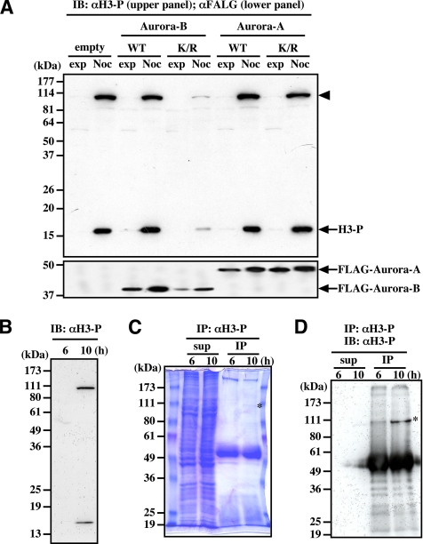
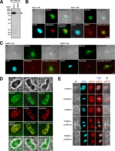
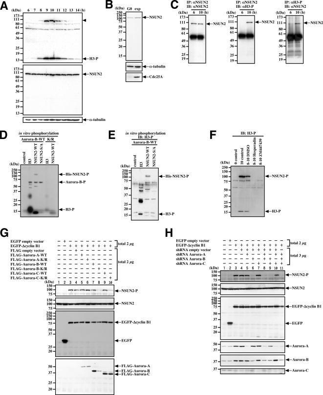
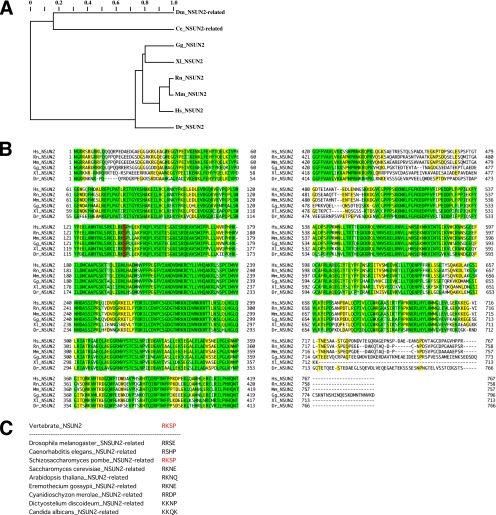
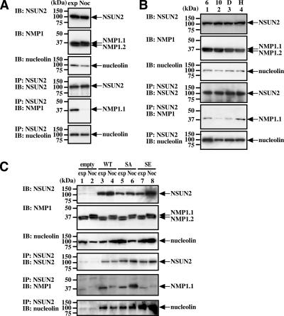
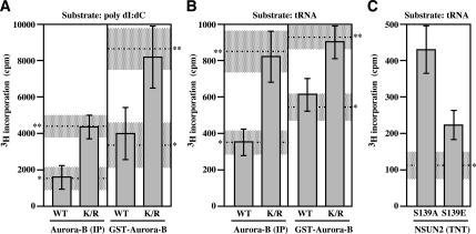
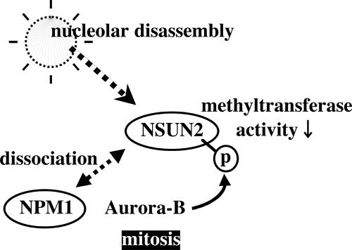
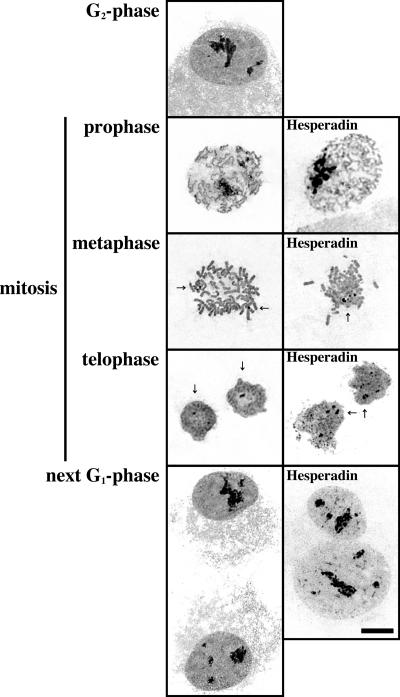
Disassembly of the nucleolus during mitosis is driven by phosphorylation of nucleolar proteins. RNA processing stops until completion of nucleolar reformation in G(1) phase. Here, we describe the RNA methyltransferase NSUN2, a novel substrate of Aurora-B that contains an NOL1/NOP2/sun domain. NSUN2 was concentrated in the nucleolus during interphase and was distributed in the perichromosome and cytoplasm during mitosis. Aurora-B phosphorylated NSUN2 at Ser139. Nucleolar proteins NPM1/nucleophosmin/B23 and nucleolin/C23 were associated with NSUN2 during interphase. In mitotic cells, association between NPM1 and NSUN2 was inhibited, but NSUN2-S139A was constitutively associated with NPM1. The Aurora inhibitor Hesperadin induced association of NSUN2 with NPM1 even in mitosis, despite the silver staining nucleolar organizer region disassembly. In vitro methylation experiments revealed that the Aurora-B-phosphorylation and the phosphorylation-mimic mutation (S139E) suppressed methyltransferase activities of NSUN2. These results indicate that Aurora-B participates to regulate the assembly of nucleolar RNA-processing machinery and the RNA methyltransferase activity of NSUN2 via phosphorylation at Ser139 during mitosis.


Figures

Figure 1.
Figure 1.
Antibody against Histone H3 phosphorylated at Ser10 binds to unknown 100-kDa protein. (A) The 100-kDa protein (arrowhead) is phosphorylated in cells treated for 18 h with nocodazole, and its phosphorylation is inhibited by the expression of Aurora-B-K/R but not Aurora-A-K/R. (B) The 100-kDa protein is phosphorylated during mitosis in synchronized cells. (C) Coomassie brilliant blue staining of the 100-kDa protein immunoprecipitated by αH3-P. Synchronized HeLa cells were collected at the indicated times, and the immunoprecipitated samples and the supernatant were separated by SDS-PAGE. (D) Immunoblot of the 100-kDa protein immunoprecipitated by αH3-P. Samples shown in C were analyzed by immunoblotting with αH3-P. In C and D, asterisks indicate the immunoprecipitated 100-kDa protein. IB, immunoblot; IP, immunoprecipitation; H3, Histone H3; WT, wild-type; K/R, kinase negative form; exp, exponentially growing; Noc, nocodazole treatment.
Figure 2.
Figure 2.
Intracellular localization of NSUN2. (A) HeLa cells express higher levels of NSUN2 (arrowhead) than NHDF cells during the exponential growth phase. (B) In interphase HeLa cells, NSUN2 is localized in the nucleoplasm and concentrated in the nucleolus. (C) In interphase NHDF cells, NSUN2 is localized in nucleoplasm and concentrated in the nucleolus. (D) In the nucleolus, NSUN2 is localized in the dense fibrillar and granular components and is not significantly associated with the fibrillar centers. (E) NSUN2 is localized in the perichromosomal and cytoplasmic regions during mitosis and is translocated into the nucleoplasm during cytokinesis. Bar, 10 μm (B and C) and 1 μm (D).
Figure 3.
Figure 3.
NSUN2 is phosphorylated during mitosis on Ser139 by Aurora-B. (A) NSUN2 is constitutively expressed and possibly phosphorylated during mitosis. The samples were collected at the indicated times in synchronized HeLa cells. The top, middle, and bottom panels show immunoblots (IB) using αH3-P, αNSUN2, and anti-α-tubulin antibody, respectively. Arrowhead indicates the phosphorylated 100-kDa protein, presumably phosphorylated by NSUN2. (B) NSUN2 is expressed even in the quiescent state. NHDF cells were cultured under exponential (exp) growth conditions or in 0.2% fetal bovine serum for 48 h (G0). (C) NSUN2 is phosphorylated during mitosis but not during interphase in synchronized HeLa cells. (D) NSUN is phosphorylated by Aurora-B in vitro. (E) NSUN2 phosphorylated in vitro by Aurora-B is recognized by αH3-P. The samples shown in D were analyzed by immunoblotting using αH3-P. (F) Aurora inhibitors Hesperadin and ZM447439 repress NSUN2 and Histone H3 phosphorylation during mitosis in synchronized HeLa cells. Eight hours after release, the cells were treated with inhibitors for 2 h. The control lysate was collected immediately after release. (G) Aurora-B-K/R and Aurora-C-K/R repress the phosphorylation of NSUN2 during mitosis in ΔCyclin B1-arrested cells. HeLa cells were transfected with the indicated pair of plasmids, and after 18 h, cells were lysed and analyzed by immunoblotting by using αH3-P, αNSUN2, anti-EGFP antibody, or anti-FLAG antibody. (H) Aurora-B-shRNA represses the phosphorylation of NSUN2 during mitosis in ΔCyclin B1-arrested cells. HeLa cells were transfected with the indicated pair of plasmids, and after treatment for 48 h with puromycin, cells were lysed and analyzed by immunoblotting αH3-P, αNSUN2, anti-EGFP antibody, or anti-Aurora-A, -B, or -C antibodies. H3, Histone H3; H3-P, phosphorylated Histone H3; NSUN2-P, phosphorylated NSUN2; Aurora-B-P, phosphorylated Aurora-B; IB, immunoblot; IP, immunoprecipitation.
Figure 4.
Figure 4.
NSUN2 is conserved among vertebrates and is distantly related to proteins in other species. (A) Tentative evolutionary tree for the vertebrate NSUN2 family and distantly related proteins in insects and nematodes. (B) Multiple sequence alignment for the vertebrate NSUN2 family. Colors indicate the extent of similarity: yellow, >60%; and green, 100%. The conserved Aurora-B phosphorylation site is indicated in red. (C) Nonconservation of the Aurora-B phosphorylation site among the NSUN2-related proteins in nonvertebrates.
Figure 5.
Figure 5.
NSUN2 associates with NPM1 and nucleolin, but this association is reduced during mitosis. (A) Association between NSUN2 and NPM1 is reduced in nocodazole-treated cells. Exponentially growing and nocodazole-treated HeLa cells were analyzed by immunoprecipitation. (B) The association between NSUN2 and NPM1 is reduced during mitosis in synchronized cells. This reduction is inhibited by Hesperadin. Eight hours after release, synchronized HeLa cells were treated for 2 h with Hesperadin or dimethyl sulfoxide (DMSO). Samples were collected at 6 (lane 1) and 10 h (lane 2) after release (after 2 h with DMSO (lane 3) or Hesperadin (lane 4)). (C) NSUN2-S139A, which mimics unphosphorylated NSUN2, is associated with NPM1 even in mitotic cells, but the association of NPM1 with NSUN2-S139E, which mimics constitutively phosphorylated NSUN2, is reduced. HeLa-NSUN2-KD cells were transfected with a plasmid for expressing NSUN2-WT, NSUN2- SA, or NSUN2-SE and were then treated for 18 h with or without nocodazole. IB, immunoblot; IP, immunoprecipitation; WT, wild-type; SA, NSUN2-S139A; SE, NSUN2-S139E; exp, exponentially growing; Noc, nocodazole treatment.
Figure 6.
Figure 6.
Phosphorylation status of NSUN2 affects its methyltransferase activities. (A) [3H]methylation of poly(dI:dC) DNA upon incubation for 30 min with purified NSUN2 after phosphorylation reaction with immunoprecipitated Aurora-B or GST-Aurora-B. (B) [3H]methylation of tRNA upon incubation for 30 min with purified NSUN2 after phosphorylation reaction with immunoprecipitated Aurora-B or GST-Aurora-B. (C) [3H]methylation of tRNA upon incubation for 5 min with TNT-produced NSUN2-S139A and NSUN2-S139E. Error bar, SD (n = 3)., basal incorporation level (shadow is SD range)., incorporation level in a reaction mixture with purified NSUN2 following MOCK phosphorylation reaction (shadow is SD range).
Figure 7.
Figure 7.
Schematic model of NSUN2 regulation by Aurora-B.
Figure 8.
Figure 8.
Aurora-B kinase activity does not affect nucleolar Ag-NOR disassembly-reassembly processes during mitosis. Ag-NOR silver staining images visualizing the nucleolus in interphase nuclei and NOR on mitotic chromosomes in Hesperadin-treated or nontreated synchronized HeLa cells. Arrow, Ag-NOR.

Similar articles

Cited by

References

    1. Adams R. R., Maiato H., Earnshaw W. C., Carmena M. Essential roles of Drosophila inner centromere protein (INCENP) and aurora B in histone H3 phosphorylation, metaphase chromosome alignment, kinetochore disjunction, and chromosome segregation. J. Cell Biol. 2001;153:865–880. - PMC - PubMed
    1. Alexandrov A., Chernyakov I., Gu W., Hiley S. L., Hughes T. R., Grayhack E. J., Phizicky E. M. Rapid tRNA decay can result from lack of nonessential modifications. Mol. Cell. 2006;21:87–96. - PubMed
    1. Andrews P. D., Knatko E., Moore W. J., Swedlow J. R. Mitotic mechanics: the auroras come into view. Curr. Opin. Cell Biol. 2003;15:672–683. - PubMed
    1. Beausoleil S. A., Jedrychowski M., Schwartz D., Elias J.E., Villen J., Li J., Cohn M. A., Cantley L. C., Gygi S. P. Large-scale characterization of HeLa cell nuclear phosphoproteins. Proc. Natl. Acad. Sci. USA. 2004;101:12130–12135. - PMC - PubMed
    1. Carmena M., Earnshaw W. C. The cellular geography of aurora kinases. Nat. Rev. Mol. Cell Biol. 2003;4:842–854. - PubMed

Publication types

MeSH terms