. 2007 Mar;18(3):1030-43.
doi: 10.1091/mbc.e06-04-0294. Epub 2007 Jan 10.

EphA4 signaling regulates blastomere adhesion in the Xenopus embryo by recruiting Pak1 to suppress Cdc42 function

Affiliations

EphA4 signaling regulates blastomere adhesion in the Xenopus embryo by recruiting Pak1 to suppress Cdc42 function

Nicolas Bisson et al. Mol Biol Cell. 2007 Mar.

Abstract

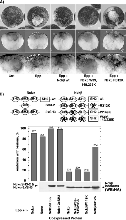
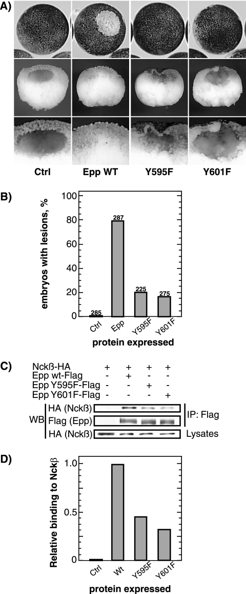
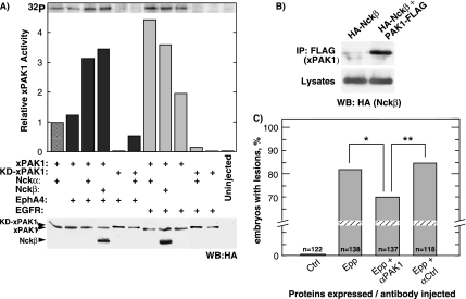
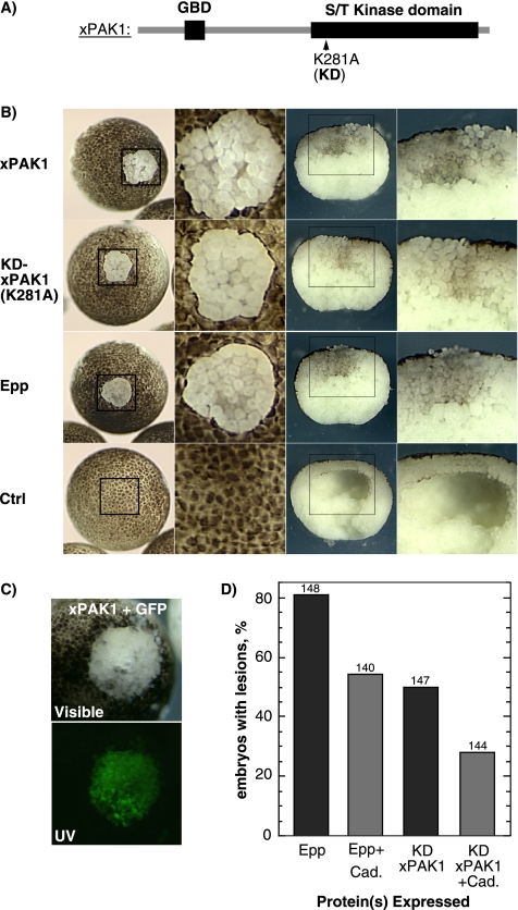
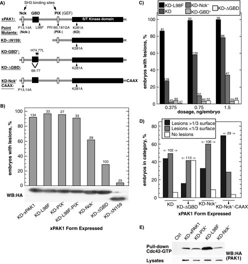
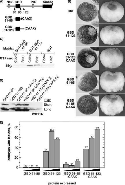
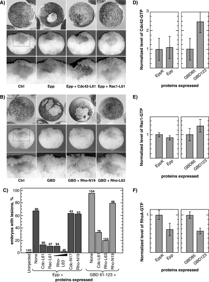
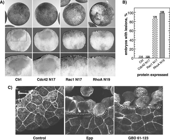
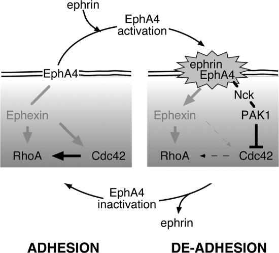
The control of cell adhesion is an important mechanism by which Eph receptors regulate cell sorting during development. Activation of EphA4 in Xenopus blastulae induces a reversible, cell autonomous loss-of-adhesion and disruption of the blastocoel roof. We show this phenotype is rescued by Nckbeta (Grb4) dependent on its interaction with EphA4. Xenopus p21(Cdc42/Rac)-activated kinase xPAK1 interacts with Nck, is activated in embryo by EphA4 in an Nck-dependent manner, and is required for EphA4-induced loss-of-adhesion. Ectopic expression of xPAK1 phenocopies EphA4 activation. This does not require the catalytic activity of xPAK1, but it does require its GTPase binding domain and is enhanced by membrane targeting. Indeed, membrane targeting of the GTPase binding domain (GBD) of xPAK1 alone is sufficient to phenocopy EphA4 loss-of-adhesion. Both EphA4 and the xPAK1-GBD down-regulate RhoA-GTP levels, and consistent with this, loss-of-adhesion can be rescued by activated Cdc42, Rac, and RhoA and can be epistatically induced by dominant-negative RhoA. Despite this, neither Cdc42 nor Rac activities are down-regulated by EphA4 activation or by the xPAK1-GBD. Together, the data suggest that EphA4 activation sequesters active Cdc42 and in this way down-regulates cell-cell adhesion. This novel signaling pathway suggests a mechanism for EphA4-guided migration.


Figures

Figure 1.
Figure 1.
Nckβ overexpression rescues the Epp loss-of-adhesion phenotype. (A) Example of a lesion caused by loss of animal pole blastomere adhesion resulting from the expression of Epp. Examples of rescue of this phenotype by the coexpression of Nckβ forms are also shown. The middle panels show corresponding manually cross-sectioned embryos, and the boxed regions are shown at higher magnification in the bottom panels. (B) The structure of Nckα and Nckβ and their mutant forms is shown above a histogram of the percentage of embryos displaying loss-of-adhesion lesions. Scoring was on the basis of visible lesions; no correction for lesion size was made. The ratio of Epp to Nck RNA injected was maintained at 1-4. The numbers above the histogram columns refer to the total number of embryos scored. The bottom panel shows the relative Nckβ and Nckα SH3-2 and 3xSH3 expression levels determined by Western analysis using an antibody against the HA epitope tag. Nckα wild type (wt) was not tagged and hence could not be detected in this way.
Figure 2.
Figure 2.
Tyrosines 595 and 601 on EphA4 are essential for the loss-of-adhesion phenotype and for Nckβ interaction. (A) Example of a lesion caused by loss of animal pole blastomere adhesion resulting from the expression of Epp and block of this phenotype by independent mutagenesis of two conserved tyrosines in the juxtamembrane domain. (B) Histogram of the percentage of embryos displaying loss-of-adhesion lesions. Scoring was on the basis of visible lesions, no correction for lesion size was made. The same amount of RNA was injected for wild-type (wt) or mutant Epp. The numbers above the histogram columns refer to the total number of embryos scored over three independent experiments. (C) Interaction of xEpp with Nckβ. HA-Nckβ and xEpp-FLAG (wt or mutants) were transfected in 293T cells, and total protein extracts were immunoprecipitated with anti-FLAG antibody, Western blotted, and probed with anti-HA antibody. (D) The data in C were quantified and are shown normalized to the wt Epp–Nckβ interaction levels.
Figure 3.
Figure 3.
xPAK1 is activated by EphA4 in embryo. (A) Assay of xPAK1 kinase activity in extracts from embryos coexpressing xPAK1 or a catalytically inactive xPAK1 (KD-xPAK1), the receptors EphA4 or hEGFR and the two Nck isoforms. Relative xPAK1 and Nckβ expression were determined by Western analysis by using an antibody to the HA epitope-tag. Nckα was not epitope tagged and so was not detected in this assay. (B) Interaction of xPAK1 with Nckβ. HA-Nckβ and xPAK1-FLAG were transfected in 293T cells, and total protein extracts were immunoprecipitated with anti-FLAG antibody, Western blotted, and probed with anti-HA antibody. (C) xPAK1 is required for loss-of-adhesion. Histogram showing the percentage of embryos displaying loss-of-adhesion lesions when injected with Epp alone or when coinjected with an affinity-purified anti-xPAK1 (αPAK1) antibody or a control antibody (αCtrl). Scoring was on the basis of visible lesions, no correction for lesion size was made. Statistical analysis showed that differences were significant; *p = 0.0199 and **p = 0.0106.
Figure 4.
Figure 4.
xPAK1 induces the loss-of-adhesion phenotype that can be suppressed by C-cadherin. (A) xPAK1 domain structure indicating the ATP binding site mutation used to inactivate the kinase domain. (B) Left panel shows examples of control embryos and embryos displaying loss of blastomere adhesion induced by xPAK1 or the KD mutant in comparison with the activated EphA4 (Epp) phenotype. The corresponding boxed regions are shown at higher magnification in the middle-left panels. The middle-right panels show manually cross-sectioned embryos, and the corresponding boxed regions are shown at higher magnification in the right panels. (C) Coexpression of xPAK1 with GFP shows that the effect of xPAK1 is restricted to the expressing cells. Visible and UV refer to incident and fluorescent light images, respectively. (D) Epp and KD-xPAK1 RNAs were coinjected with or without C-cadherin RNA, and loss-of-adhesion lesions was scored. The ratio of Epp or xPAK1 to cadherin RNA was maintained at 1-4. Scoring was on the basis of visible lesions; no correction for lesion size was made. The numbers above the histogram columns refer to the total number of embryos scored.
Figure 5.
Figure 5.
The xPAK1 loss-of-adhesion phenotype requires the GBD and is enhanced by the Nck binding site and by CAAX targeted membrane recruitment. (A) The structure of the xPAK1 mutants used. Amino acid mutations and the extent of deletion mutations are indicated as is the C-terminal CAAX extension. (B) Each xPAK1 mutant was injected and loss-of-adhesion lesions were scored. RNA injections were adjusted to give a high penetrance of the KD-xPAK1–induced phenotype, and this same amount of each mutant RNA was then injected. Bottom panel shows relative protein expression levels as determined by Western analysis by using an antibody to the HA epitope-tag. Two regions of the same Western analysis are shown. (C) Dose–response relationship for increasing amounts of injected KD-L98F, KD, KD-Nck, KD-GBD, and KD-ΔGBD-xPAK1 RNAs. Scoring was regardless of lesion size. (D) Comparison of lesion size. Equal RNA amounts of each mutant were injected, and the sizes of loss-of-adhesion lesions were categorized relative to the total animal pole surface. The numbers above the histogram columns refer to the total number of embryos scored. (E) KD-L98F xPAK1 displays an increased affinity for GTP-Cdc42. HA-tagged xPAK1 constructs were expressed in embryo, whole extracts applied to Sepharose-bound GST-Cdc42 fusion protein precharged with GTP, and revealed by Western blotting by using the HA epitope tag.
Figure 6.
Figure 6.
The functional GBD of xPAK1 is sufficient to induce the loss-of-adhesion phenotype, but its function is enhanced by membrane targeting. (A) The structure of the GBD constructs (xPAK1 a.a. 61-85 and a.a. 61-123). (B) Phenotype of the GBD-induced lesions, left panels show external animal pole views, and right panels show sections through the lesions. (C) Binding of in vitro-translated GTP-charged wild type Cdc42 and Rac1 GTPases to the GST-immobilized xPAK1 GBDs 61-85 and 61-123. (D) Western analysis of expression levels of the GBD constructs using an antibody to the common HA epitope-tag (h embryos in 1× MMR and l embryos in 0.1× MMR). Two exposures of the same analysis are shown to increase the visible dynamic range. (E) Statistical analysis of lesion penetrance. Embryos were injected in parallel with 70, 200, or 400 pg of GBD 61-123 and GBD 61-123-CAAX RNAs. Embryos displaying obvious lesions, irrespective of size, were scored. Total number of embryos analyzed is shown above each column. In B and D, each embryo was injected with 200 pg of mutant RNA.
Figure 7.
Figure 7.
Activated Cdc42, Rac1, and RhoA rescue EphA4 induced loss-of-adhesion. (A) Epp RNA was coinjected with activated Cdc42 or Rac1 GTPases RNA (ratio 1:3) at the two-cell stage. Lower panels show corresponding sections of embryos in top panels, and corresponding boxed regions are shown at higher magnification in the bottom panels. (B) As in A, but the xPAK1-GBD 61-123 was coinjected either alone or with dominant-negative (N19) or activated RhoA (L63) (ratio 1:2). Bottom panels show sections of the corresponding embryos, and corresponding boxed regions are shown at higher magnification in the bottom panels. Typical examples of resulting embryos are shown. See Supplemental Figure 1 for sample images of dominant-negative GTPase coinjection. (C) Quantitation of Epp and GBD rescue with active and dominant-negative forms of Cdc42, Rac1, and RhoA. The numbers above the histogram columns refer the total number of embryos scored. (D) Levels of active (GTP-bound) Cdc42 in embryos expressing Epp or EppK (kinase dead Epp that does not give the loss-of-adhesion phenotype) and xPAK1 GBD constructs. Active Cdc42 levels were measured by pull-down with the xPAK1 GBD. The average of three experiments is shown. (E) Levels of active (GTP-bound) Rac1, measured as in D. The average of two experiments is shown. (F) Levels of active (GTP-bound) RhoA in embryos expressing Epp or EppK or xPAK1 GBD constructs. Active RhoA levels were measured by pull-down with the rhotekin GBD. The average of two experiments is shown.
Figure 8.
Figure 8.
DN-RhoA and DN-Rac1 also induce loss-of-adhesion. (A) Phenotypes of the DN-Cdc42 (N17), DN-Rac1 (N17), and DN-RhoA (N19) expression in embryo. Top panels show external animal pole views, middle panels show sections through the lesions, and corresponding boxed regions are shown at higher magnification in the bottom panels. Typical examples of resulting embryos are shown. (B) Histogram of the percentage of embryos displaying loss-of-adhesion lesions. Scoring was on the basis of visible lesions; no correction for lesion size was made. The same amount of GTPases was injected for all mutants. The numbers above the histogram columns refer to the total number of embryos scored over two independent experiments. For DN-Rac1, the shaded bar represents the penetrance of the distinct phenotype shown in A. (C) Confocal imaging of the actin cytoskeleton in a phalloidin-stained control embryo and in embryos undergoing Epp and xPAK1-GBD (61-123) loss-of-adhesion. The boundary of the loss-of-adhesion lesion is indicated by a dotted line.
Figure 9.
Figure 9.
Schematic diagram describing the probable mechanisms of induction of the loss-of-adhesion lesions by EphA4. Pointed arrowed lines show activating and blunt ended lines inhibitory signaling and line density indicates signaling strength.

Similar articles

Cited by

References

    1. Adler C. E., Miyoshi-Akiyama T., Aleman L. M., Tanaka M., Smith J. M., Mayer B. J. Abl family kinases and Cbl cooperate with the Nck adaptor to modulate Xenopus development. J. Biol. Chem. 2000;275:36472–36478. - PubMed
    1. Bagrodia S., Taylor S. J., Jordon K. A., Van Aelst L., Cerione R. A. A novel regulator of p21-activated kinases. J. Biol. Chem. 1998;273:23633–23636. - PubMed
    1. Becker E., Huynh-Do U., Holland S., Pawson T., Daniel T. O., Skolnik E. Y. Nck-interacting Ste20 kinase couples Eph receptors to c-Jun N-terminal kinase and integrin activation. Mol. Cell. Biol. 2000;20:1537–1545. - PMC - PubMed
    1. Benard V., Bohl B. P., Bokoch G. M. Characterization of rac and cdc42 activation in chemoattractant-stimulated human neutrophils using a novel assay for active GTPases. J. Biol. Chem. 1999;274:13198–13204. - PubMed
    1. Bisson N., Islam N., Poitras L., Jean S., Bresnick A. R., Moss T. The catalytic domain of xPAK1 is sufficient to induce myosin II dependent in vivo cell fragmentation independently of other apoptotic events. Dev. Biol. 2003;263:264–281. - PubMed

Publication types

MeSH terms