NO and CO differentially activate soluble guanylyl cyclase via a heme pivot-bend mechanism
- PMID: 17215864
- PMCID: PMC1783457
- DOI: 10.1038/sj.emboj.7601521
NO and CO differentially activate soluble guanylyl cyclase via a heme pivot-bend mechanism
Abstract
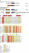
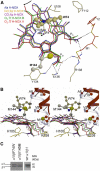
Diatomic ligand discrimination by soluble guanylyl cyclase (sGC) is paramount to cardiovascular homeostasis and neuronal signaling. Nitric oxide (NO) stimulates sGC activity 200-fold compared with only four-fold by carbon monoxide (CO). The molecular details of ligand discrimination and differential response to NO and CO are not well understood. These ligands are sensed by the heme domain of sGC, which belongs to the heme nitric oxide oxygen (H-NOX) domain family, also evolutionarily conserved in prokaryotes. Here we report crystal structures of the free, NO-bound, and CO-bound H-NOX domains of a cyanobacterial homolog. These structures and complementary mutational analysis in sGC reveal a molecular ruler mechanism that allows sGC to favor NO over CO while excluding oxygen, concomitant to signaling that exploits differential heme pivoting and heme bending. The heme thereby serves as a flexing wedge, allowing the N-terminal subdomain of H-NOX to shift concurrent with the transition of the six- to five-coordinated NO-bound state upon sGC activation. This transition can be modulated by mutations at sGC residues 74 and 145 and corresponding residues in the cyanobacterial H-NOX homolog.
Figures


Similar articles
-
Probing domain interactions in soluble guanylate cyclase.Biochemistry. 2011 May 24;50(20):4281-90. doi: 10.1021/bi200341b. Epub 2011 May 3. Biochemistry. 2011. PMID: 21491957 Free PMC article.
-
Aspartate 102 in the heme domain of soluble guanylyl cyclase has a key role in NO activation.Biochemistry. 2011 May 24;50(20):4291-7. doi: 10.1021/bi2004087. Epub 2011 May 2. Biochemistry. 2011. PMID: 21491881 Free PMC article.
-
Molecular model of a soluble guanylyl cyclase fragment determined by small-angle X-ray scattering and chemical cross-linking.Biochemistry. 2013 Mar 5;52(9):1568-82. doi: 10.1021/bi301570m. Epub 2013 Feb 15. Biochemistry. 2013. PMID: 23363317 Free PMC article.
-
How do heme-protein sensors exclude oxygen? Lessons learned from cytochrome c', Nostoc puntiforme heme nitric oxide/oxygen-binding domain, and soluble guanylyl cyclase.Antioxid Redox Signal. 2012 Nov 1;17(9):1246-63. doi: 10.1089/ars.2012.4564. Epub 2012 Apr 10. Antioxid Redox Signal. 2012. PMID: 22356101 Free PMC article. Review.
-
Structure and Activation of Soluble Guanylyl Cyclase, the Nitric Oxide Sensor.Antioxid Redox Signal. 2017 Jan 20;26(3):107-121. doi: 10.1089/ars.2016.6693. Epub 2016 Apr 26. Antioxid Redox Signal. 2017. PMID: 26979942 Free PMC article. Review.
Cited by
-
Tunnels modulate ligand flux in a heme nitric oxide/oxygen binding (H-NOX) domain.Proc Natl Acad Sci U S A. 2011 Oct 25;108(43):E881-9. doi: 10.1073/pnas.1114038108. Epub 2011 Oct 12. Proc Natl Acad Sci U S A. 2011. PMID: 21997213 Free PMC article.
-
Cryo-EM density map fitting driven in-silico structure of human soluble guanylate cyclase (hsGC) reveals functional aspects of inter-domain cross talk upon NO binding.J Mol Graph Model. 2019 Jul;90:109-119. doi: 10.1016/j.jmgm.2019.04.009. Epub 2019 Apr 24. J Mol Graph Model. 2019. PMID: 31055154 Free PMC article.
-
From synaptically localized to volume transmission by nitric oxide.J Physiol. 2016 Jan 1;594(1):9-18. doi: 10.1113/JP270297. Epub 2015 Nov 18. J Physiol. 2016. PMID: 26486504 Free PMC article. Review.
-
Structural insights into the role of iron-histidine bond cleavage in nitric oxide-induced activation of H-NOX gas sensor proteins.Proc Natl Acad Sci U S A. 2014 Oct 7;111(40):E4156-64. doi: 10.1073/pnas.1416936111. Epub 2014 Sep 24. Proc Natl Acad Sci U S A. 2014. PMID: 25253889 Free PMC article.
-
Inhibitors of NLRP3 Inflammasome Formation: A Cardioprotective Role for the Gasotransmitters Carbon Monoxide, Nitric Oxide, and Hydrogen Sulphide in Acute Myocardial Infarction.Int J Mol Sci. 2024 Aug 26;25(17):9247. doi: 10.3390/ijms25179247. Int J Mol Sci. 2024. PMID: 39273196 Free PMC article. Review.
References
-
- Ascenzi P, Bocedi A, Leoni L, Visca P, Zennaro E, Milani M, Bolognesi M (2004) CO sniffing through heme-based sensor proteins. IUBMB Life 56: 309–315 - PubMed
-
- Behrends S (2003) Drugs that activate specific nitric oxide sensitive guanylyl cyclase isoforms independent of nitric oxide release. Curr Med Chem 10: 291–301 - PubMed
-
- Boehning D, Snyder SH (2003) Novel neural modulators. Annu Rev Neurosci 26: 105–131 - PubMed
-
- Boon EM, Davis JH, Tran R, Karow DS, Huang SH, Pan D, Miazgowicz MM, Mathies RA, Marletta MA (2006) Nitric oxide binding to prokaryotic homologs of the soluble guanylate cyclase beta 1 H-nox domain. J Biol Chem 281: 21892–21902 - PubMed
Publication types
MeSH terms
Substances
Grants and funding
LinkOut - more resources
Full Text Sources

