. 2007 Jan 24;26(2):623-33.
doi: 10.1038/sj.emboj.7601500. Epub 2007 Jan 11.

Structural insights into the innate immune recognition specificities of L- and H-ficolins

Affiliations

Structural insights into the innate immune recognition specificities of L- and H-ficolins

Virginie Garlatti et al. EMBO J. .

Abstract

Innate immunity relies critically upon the ability of a few pattern recognition molecules to sense molecular markers on pathogens, but little is known about these interactions at the atomic level. Human L- and H-ficolins are soluble oligomeric defence proteins with lectin-like activity, assembled from collagen fibers prolonged by fibrinogen-like recognition domains. The X-ray structures of their trimeric recognition domains, alone and in complex with various ligands, have been solved to resolutions up to 1.95 and 1.7 A, respectively. Both domains have three-lobed structures with clefts separating the distal parts of the protomers. Ca(2+) ions are found at sites homologous to those described for tachylectin 5A (TL5A), an invertebrate lectin. Outer binding sites (S1) homologous to the GlcNAc-binding pocket of TL5A are present in the ficolins but show different structures and specificities. In L-ficolin, three additional binding sites (S2-S4) surround the cleft. Together, they define an unpredicted continuous recognition surface able to sense various acetylated and neutral carbohydrate markers in the context of extended polysaccharides such as 1,3-beta-D-glucan, as found on microbial or apoptotic surfaces.


Figures

Figure 1
Figure 1
Homotrimeric structure of the recognition domains of human L- and H-ficolins and location of their binding site(s). (A, B) L-ficolin structure seen from the target binding surface (bottom view) and on a perpendicular side view. (C, D) Corresponding bottom and side views of the H-ficolin structure. The side chains of the binding site residues are displayed as ball and sticks and colored green (S1), red (S2), black (S3), and orange (S4). To enhance clarity of the side view, only one of each representative binding sites is shown on the L-ficolin trimer. N and C indicate the N- and C-terminal ends of each protomer. Ca2+ ions are represented as golden spheres. Figure generated using MOLSCRIPT (Kraulis, 1991).
Figure 2
Figure 2
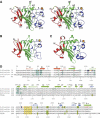
Structure of the fibrinogen-like protomers. (A) Stereo view of the L-ficolin protomer. (B, C) Comparative views of the H-ficolin and TL5A protomers in the same orientation. Domains A, B, and P are colored red, green, and blue, respectively. Disulfide bonds are shown in yellow. N and C indicate the N- and C-terminal ends. Figure generated using MOLSCRIPT. (D) Sequence alignment of the fibrinogen-like domains of L-ficolin, M-ficolin, H-ficolin, and TL5A. Numbers on top and below refer to the L-ficolin and TL5A sequences, respectively. Secondary structure elements are shown in red (A domain), green (B domain), and blue (P domain). The binding site residues are colored using the same code as in Figure 1: green (S1), bold red (S2), black (S3), and bold orange (S4). Residues involved in the inter-subunit interfaces are colored cyan, variable ones being boxed. Residues involved in Ca2+ binding in L-ficolin, H- ficolin, and TL5A are displayed in a yellow box.
Figure 3
Figure 3
Comparative views of the S1 binding site in H-ficolin, L-ficolin, and TL5A. The side chains of the residues defining S1 are colored green and the ligands are displayed in yellow. (A) D-Fucose bound to H-ficolin. (B) GlcNAc bound to TL5A. (C) The terminal mannose of the oligosaccharide chain from a neighboring molecule positioned in site S1 of L-ficolin. On the left, the two proximal GlcNAc residues of the chain interacting on the edge of the binding site.
Figure 4
Figure 4
The novel binding sites identified in L-ficolin. (A–C) Interaction of galactose, GlcNAc, and N-acetylcysteine (CysNAc) in site S2. The interaction of galactose O6 with D111, at the back of the binding pocket, is not visible on this orientation. (D) Detailed view of the interaction of GalNAc in S3. (E) Binding of acetylcholine to site S3. (F) Superimposition of different acetylated ligands bound to site S3. Three molecules have been selected for clarity purposes, showing both medium and extreme orientations. The common acetyl group interaction is highlighted with dashed lines. Acetylcholine, GalNAc, and ManNAc are shown in yellow, green, and pink, respectively. A more extensive superimposition of the different acetylated molecules bound to site S3 is provided as Supplementary data. (G) Interaction of the first three glucose units of 1,3-β-D-glucan with residues from sites S3 (black) and S4 (orange). (H) A different view of the binding of 1,3-β-D-glucan, showing interaction of Glc3 and Glc4 with residues from sites S3 (black) and S4 (orange). Polar interactions are represented by dashed red lines. Water molecules are shown as red dots and the calcium ion is represented by a yellow sphere.
Figure 5
Figure 5
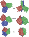
Comparative views of the trimeric recognition domains of MBL, L-ficolin, and C1q. Space-filling bottom views (left) and side views (right) are shown. Ca2+ ions are represented as yellow spheres. Figure generated using GRASP (Nicholls et al, 1991).

References

    1. Adachi Y, Ishii T, Ikeda Y, Hoshino A, Tamura H, Aketagawa J, Tanaka S, Ohno N (2004) Characterization of beta-glucan recognition site on C-type lectin, dectin 1. Infect Immun 72: 4159–4171 - PMC - PubMed
    1. Aoyagi Y, Adderson EE, Min JG, Matsushita M, Fujita T, Takahashi S, Okuwaki Y, Bohnsack JF (2005) Role of L-ficolin/mannose-binding lectin associated serine protease complexes in the opsonophagocytosis of type III group B streptococci. J Immunol 174: 418–425 - PubMed
    1. Bilyy RO, Antonyuk VO, Stoika RS (2004) Cytochemical study of role of α-D-mannose- and β-D-galactose-containing glycoproteins in apoptosis. J Mol Histol 35: 829–838 - PubMed
    1. Brünger AT, Adams PD, Clore GM, DeLano WL, Gros P, Grosse-Kunstleve RW, Jiang JS, Kuszewski J, Nilges M, Pannu NS, Read RJ, Rice LM, Simonson T, Warren GL (1998) Crystallography & NMR system: a new software suite for macromolecular structure determination. Acta Crystallogr D 54: 905–921 - PubMed
    1. Chang CI, Chelliah Y, Borek D, Mengin-Lecreulx D, Deisenhofer J (2006) Structure of tracheal cytotoxin in complex with a heterodimeric pattern-recognition receptor. Science 311: 1761–1764 - PubMed

Publication types

MeSH terms