Comparative Study
. 2006 Dec;8(12):1042-54.
doi: 10.1593/neo.06568.

SAG/ROC-SCF beta-TrCP E3 ubiquitin ligase promotes pro-caspase-3 degradation as a mechanism of apoptosis protection

Affiliations
Comparative Study

SAG/ROC-SCF beta-TrCP E3 ubiquitin ligase promotes pro-caspase-3 degradation as a mechanism of apoptosis protection

Mingjia Tan et al. Neoplasia. 2006 Dec.

Abstract

Skp1-cullin-F-box protein (SCF) is a multicomponent E3 ubiquitin (Ub) ligase that ubiquitinates a number of important biologic molecules such as p27, beta-catenin, and IkappaB for proteasomal degradation, thus regulating cell proliferation and survival. One SCF component, SAG/ROC2/Rbx2/Hrt2, a RING finger protein, was first identified as a redox-inducible protein, which, when overexpressed, inhibited apoptosis both in vitro and in vivo. We report here that sensitive to apoptosis gene (SAG), as well as its family member ROC1/Rbx1, bound to the proinactive form of caspase-3 (pro-caspase-3). Binding was likely mediated through F-box protein, beta-transducin repeat-containing protein (beta-TrCP), which binds to the first 38 amino acids of pro-caspase-3. Importantly, beta-TrCP1 expression significantly shortened the protein half-life of pro-caspase-3, whereas expression of a dominant-negative beta-TrCP1 mutant with the F-box domain deleted extended it. An in vitro ubiquitination assay showed that SAG/ROC-SCF(beta-TrCP) promoted ubiquitination of pro-caspase-3. Furthermore, endogenous levels of pro-caspase-3 were decreased by overexpression of SAG/ROC-SCF(beta-TrCP) E3 Ub ligases, but increased on siRNA silencing of SAG, regulator of cullin-1 (ROC1), or beta-TrCPs, leading to increased apoptosis by etoposide and TNF-related apoptosis-inducing ligand through increased activation of caspase-3. Thus, pro-caspase-3 appears to be a substrate of SAG/ROC-SCF(beta-TrCP) E3 Ub ligase, which protects cells from apoptosis through increased apoptosis threshold by reducing the basal level of pro-caspase-3.


Figures

Figure 1
Figure 1
SAG or ROC1 bound to endogenous pro-caspase-3 in 293 cells. Human 293 cells were transiently transfected with pcDNA3 vector control, FLAG-tagged SAG (A), or HA-tagged ROC1 (B), using Lipofectamine 2000. Cell lysates were prepared after transfection and subjected to IP with anti-caspase-3 antibody or control IgG, as indicated after preclearing with protein A/G-plus agarose beads, followed by immunoblotting using anti-FLAG antibody or anti-HA antibody, respectively. The bottom panel shows direct immunoblotting using whole-cell extract with anti-FLAG or anti-HA antibody, respectively, to show that transfected SAG or ROC1 was expressed in 293 cells. (C) SAG-β-TrCP2 binding. Human 293 cells were transiently cotransfected with HA-β-TrCP2 and FLAG-SAG or pcDNA3 vector control. After transfection, cell lysates were prepared and subjected to IP using anti-FLAG antibody or control IgG, as indicated after preclearing with protein A/G-plus agarose beads, followed by immunoblotting with anti-HA antibody (top). Expression of transfected β-TrCP2 (middle) and SAG (bottom) was detected by direct immunoblotting of whole-cell extract. (D) SAG-β-TrCP1 binding. Human 293 cells were cotransfected with FLAG-β-TrCP1 and HA-SAG or pcDNA3 vector control. Cell lysates were prepared and subjected to IP using anti-FLAG antibody or control IgG, as indicated after preclearing with protein A/G-plus agarose beads, followed by immunoblotting using anti-HA antibody (top). Expression of transfected β-TrCP1 (middle) and SAG (bottom) was detected by direct immunoblotting of whole-cell extract.
Figure 2
Figure 2
β-TrCPs bound to pro-caspase-3 require prodomain. (A) β-TrCP2 bound to endogenous pro-caspase-3. Human 293 cells were transiently transfected with HA-β-TrCP2 or pcDNA3 vector control. Cell lysates were prepared and subjected to IP using anti-caspase-3 antibody or preimmune IgG, as indicated after preclearing with protein A/G-plus agarose beads, followed by immunoblotting using anti-HA antibody (top panel). A direct immunoblotting analysis of whole-cell extract was performed using anti-HA antibody to show β-TrCP2 expression (bottom). (B) β-TrCP1 bound to endogenous pro-caspase-3. Human 293 cells were transiently transfected with pcDNA3 vector control (lane 1), FLAG-β-TrCP1 (lane 2), and FLAG-β-TrCP1δF (lane 3). Cell lysates were prepared and subjected to IP using anti-caspase-3 antibody followed by Western blot analysis with anti-FLAG antibody (top), or whole-cell extracts were directly subjected to Western blot analysis to show expression of FLAG-β-TrCP1 and FLAG-β-TrCP1ΔF (bottom). Requirement of prodomain for β-TrCP-pro-caspase-3 binding. (C) Bar graphic presentation of pro-caspase-3 mutants. (D–F) Human 293 cells were cotransiently transfected with HA-β-TrCP2 in combination with WT pro-caspase-3 or its deletion mutants, along with the vector control. Thirty-eight hours posttransfection, cells were harvested and subjected to Western blot analysis (D and E) using indicated antibodies or IP, followed by Western blot analysis using myc antibody (F). NB, nonspecific band.
Figure 3
Figure 3
β-TrCPs shortened the protein half-life of pro-caspase-3. (A) β-TrCP1 induced a dose-dependent reduction of pro-caspase-3. Pro-caspase-3-null MCF7 cells were cotransfected with FLAG-pro-caspase-3 and an increasing amount of β-TrCP1. Thirty-eight hours posttransfection, cell lysates were prepared and subjected to direct Western blot analysis using anti-caspase-3 antibody, as well as anti-actin antibody as loading control. (B and C) β-TrCP1 shortened—but its dominant-negative mutant extended—the pro-caspase-3 half-life. MCF7 cells were transiently cotransfected with FLAG-pro-caspase-3, along with pcDNA3 vector control, β-TrCP1, or β-TrCP1ΔF, as indicated. Twenty-four hours posttransfection, cells were treated with CHX (10 µg/ml) for indicated time periods up to 7 hours and harvested. Cell lysates were prepared and subjected to Western blot analysis with anti-FLAG antibody or anti-β-actin antibody as loading control. Densitometric quantification of pro-caspase-3 levels was shown in (C), with value at 0 hour after CHX treatment arbitrarily set to 1, on which the fold change was calculated.
Figure 4
Figure 4
Pro-caspase-3 ubiquitination by SAG/ROC-SCF E3 Ub ligase. (A) ROC1-SCFβ-TrCP E3 ligase. Bacterially expressed and purified FLAG-tagged pro-caspase-3 was combined with Ub, Uba1 (E1), UbcH3 (E2), and E3, ROC1-SCFβTrCP1, in which each component was individually expressed in baculovirus and copurified as a four-component complex, as described in Materials and Methods section. Omission of some components was carried out as indicated. Samples were incubated in the presence of ATP buffer for 1.5 hours at 37°C, run on a 10% to a 15% gradient SDS-PAGE, and blotted with anti-pro-caspase-3 antibody. *Nonspecific band. HC indicates the heavy chain of IgG. (B) SAG-SCFβ-TrCP. Bacterially purified FLAG-tagged pro-caspase-3 and GST-SAG were combined with Ub, Uba1 (E1), Ubc3 (E2), and SCFβTrCP1, in which each component was individually expressed in baculovirus and copurified as a three-component complex, as described in Materials and Methods section. Omission of pro-caspase-3 on the right lane was indicated. Samples were incubated in the presence of ATP buffer for 1.5 hours at 37°C, run on a 10% SDS-PAGE, and blotted with anti/pro-caspase-3 antibody. HC, heavy chain from FLAG affinity purification. *Nonspecific signals.
Figure 5
Figure 5
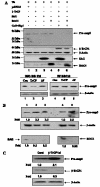
Pro-caspase-3 is a substrate of SAG/ROC1-SCFβ-TrCP E3 ligase. (A) SAG/ROC-SCFβ-TrCP E3 Ub ligase promoted the degradation of endogenous pro-caspase-3. Top panel: Human 293 cells were transiently transfected with Ub, along with the pcDNA vector control (lane 1), FLAG-β-TrCP (lane 2), FLAG-SAG (lane 3), HA-ROC1 (lane 4), all four components of SAG-SCF E3 Ub ligase (SAG-SCF, SAG + β-TrCP1 + Cul1 + Skp1, lane 5), or all four components of ROC1-SCF E3 ligase (ROC1-SCF; lane 6), respectively. Cell lysates were prepared 38 hours posttransfection and subjected to Western blot analysis with anti-caspase-3 antibody. The blots were stripped and reprobed with antibodies against FLAG tag for the expression of β-TrCP1 (panel 2), SAG (panel 3), HA tag for ROC1 expression (panel 4), and β-actin (panel 2) for loading control. Bottom panel: Proteasomal-dependent degradation of pro-caspase-3 by SAG-SCFβ-TrCP1 E3 ligase. Human 293 cells were transiently transfected with Ub, along with the pcDNA vector control (lanes 1 and 4), SAG-SCFβ-TrCP1 (lanes 2 and 5), or SAG-SCFβ-TrCP1ΔF (a substitution of wild-type β-TrCP1 with its F-box-deleted mutant; lanes 3 and 6). Twenty-four hours posttransfection, cells were either left untreated (left panel) or treated with 10 µM MG132 (right panel) for 16 hours. Cell lysates were then prepared and subjected to Western blot analysis, with antibodies against caspase-3 or actin as loading control. (B) siRNA silencing of SAG or ROC1 increased the endogenous level of pro-caspase-3. HeLa cells were transfected with plasmid-based hairpin RNA designed to silence SAG or ROC1: psiSAG-1 (lane 2) and psiSAG-2 (lane 3), as well as psiROC-1 (lane 5), along with control psiCont (lanes 1 and 4). Thirty-eight hours posttransfection, cell lysates were prepared and subjected to Western blot analysis using antibodies against caspase-3 (top panel), actin (middle panel) or SAG (lanes 1–3, bottom panel), or ROC1 (lanes 4 and 5, bottom panel). The value of densitometric quantification after normalization with β-actin for equal protein loading was shown on the bottom of each panel, with control number setting at 1, on which the fold change was calculated. (C) siRNA silencing of β-TrCP1 increased the endogenous level of pro-caspase-3. HeLa cells were transfected with plasmid-based hairpin RNA designed to silence β-TrCP1: β-TrCP1-si, along with control psiCont (Cont). Thirty-eight hours posttransfection, cell lysates were prepared and subjected to Western blot analysis using antibody against caspase-3 (top panel), β-TrCP1 (middle), or β-actin (bottom). The value of densitometric quantification after normalization with β-actin for equal protein loading was shown on the bottom of each panel, with control number setting at 1 on which the fold change was calculated.
Figure 6
Figure 6
Silencing of SCF components results in caspase-3 activation and apoptosis induction. (A) Silencing of SAG, ROC1, or β-TrCPs increased caspase-3 activity upon activation and (B) cell death induced by anticancer agents. HeLa cells were transfected with psiSAG-2, psiROC-1, or β-TrCP-si alone, along with psiCont plasmid. Thirty-six hours posttransfection, cells were treated with either DMSO or etoposide (25 µM) for 24 hours, or TRAIL (50 ng/ml) for 8 hours, followed by caspase-3 activity assay (A) or trypan blue staining assay for cell viability (B). The results were presented as percent control (mean ± SEM) from three independent experiments. Paired Student's t test was used to define statistically significant levels at *P < .05 or **P < .01.

Similar articles

Cited by

References

    1. Thompson CB. Apoptosis in the pathogenesis and treatment of disease. Science. 1995;267(5203):1456–1462. - PubMed
    1. Wyllie AH, Kerr JF, Currie AR. Cell death: the significance of apoptosis. Int Rev Cytol. 1980;68:251–306. - PubMed
    1. Hengartner MO. The biochemistry of apoptosis. Nature. 2000;407(6805):770–776. - PubMed
    1. Earnshaw WC, Martins LM, Kaufmann SH. Mammalian caspases: structure, activation, substrates, and functions during apoptosis. Annu Rev Biochem. 1999;68:383–424. - PubMed
    1. Cryns V, Yuan J. Proteases to die for. Genes Dev. 1998;12(11):1551–1570. - PubMed

Publication types

LinkOut - more resources