. 2007 Jan 12;128(1):141-56.
doi: 10.1016/j.cell.2006.11.040.

Ubiquitination regulates PTEN nuclear import and tumor suppression

Affiliations

Ubiquitination regulates PTEN nuclear import and tumor suppression

Lloyd C Trotman et al. Cell. .

Abstract

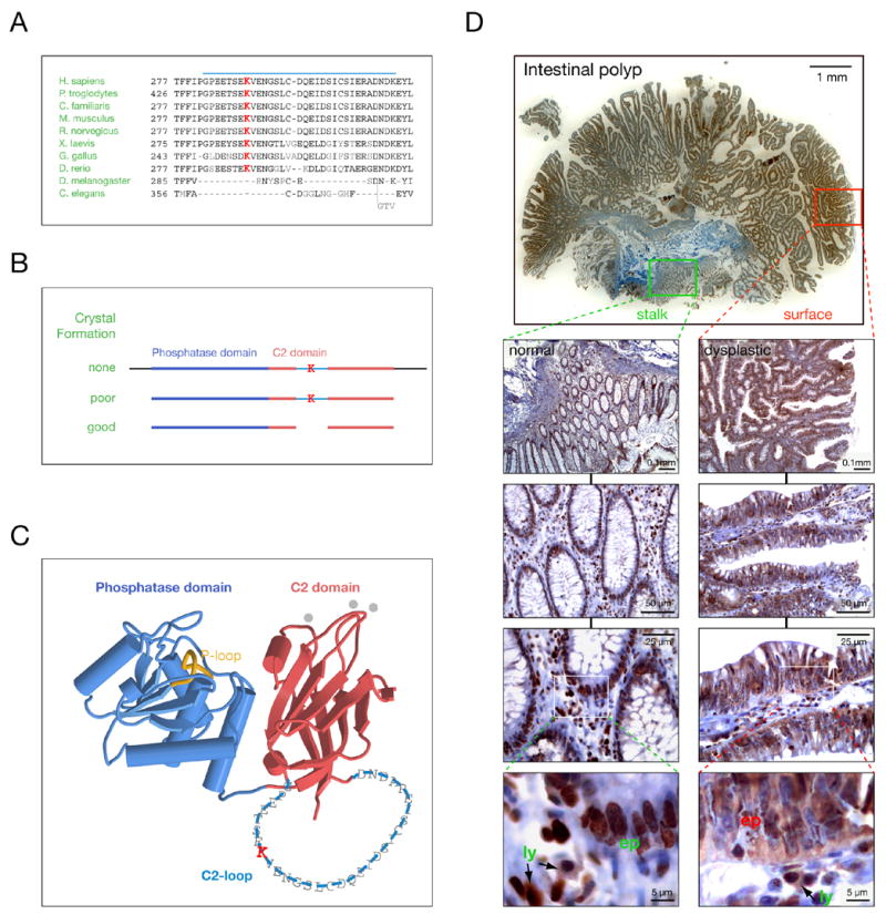
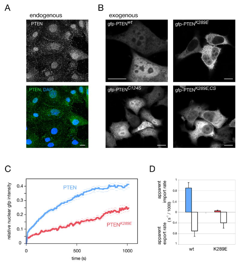
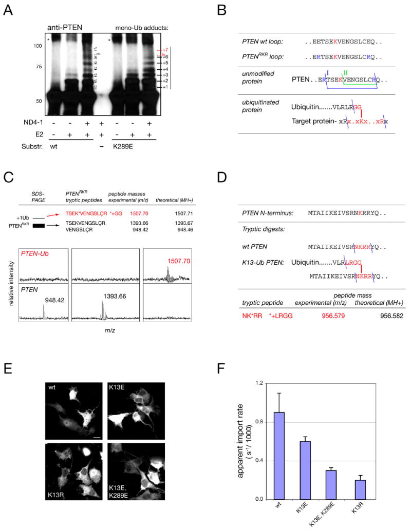
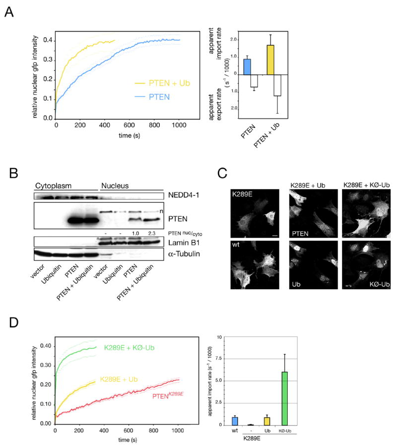
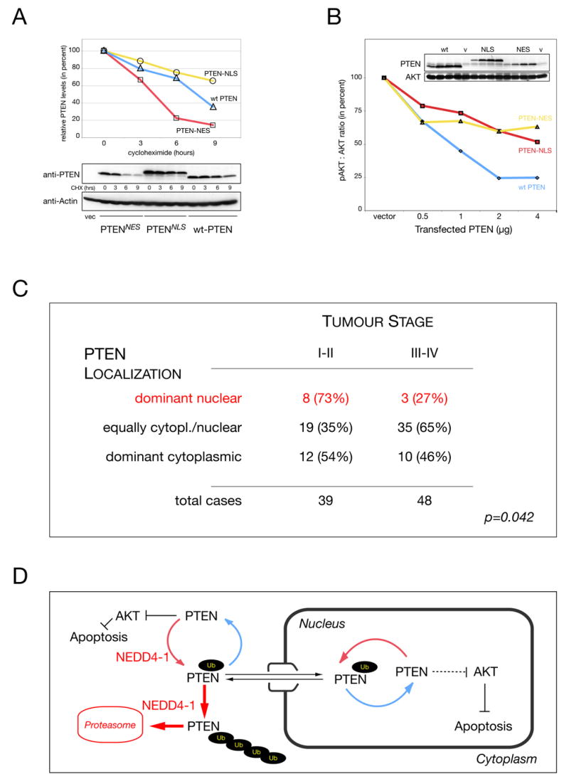
The PTEN tumor suppressor is frequently affected in cancer cells, and inherited PTEN mutation causes cancer-susceptibility conditions such as Cowden syndrome. PTEN acts as a plasma-membrane lipid-phosphatase antagonizing the phosphoinositide 3-kinase/AKT cell survival pathway. However, PTEN is also found in cell nuclei, but mechanism, function, and relevance of nuclear localization remain unclear. We show that nuclear PTEN is essential for tumor suppression and that PTEN nuclear import is mediated by its monoubiquitination. A lysine mutant of PTEN, K289E associated with Cowden syndrome, retains catalytic activity but fails to accumulate in nuclei of patient tissue due to an import defect. We identify this and another lysine residue as major monoubiquitination sites essential for PTEN import. While nuclear PTEN is stable, polyubiquitination leads to its degradation in the cytoplasm. Thus, we identify cancer-associated mutations of PTEN that target its posttranslational modification and demonstrate how a discrete molecular mechanism dictates tumor progression by differentiating between degradation and protection of PTEN.


Figures

Figure 1
Figure 1. The PTENK289E Cowden mutant shows nuclear exclusion in patient samples
(A) Sequence alignment of the PTEN C2-loop (blue bar) from various species shows K289 conservation in vertebrates (highlighted in red). (B) PTEN cleavage requirements for crystallization reveal that the C2-loop does not contribute to structural integrity (K denotes K289). (C) Localization and size of the C2-loop within the PTEN structure (single amino acid code over backbone). Note that the C2-loop faces away from the cell membrane (top) and membrane-interacting loops of the C2-domain (grey dots). (D) Intestinal polyp from a PTENwt/K289E Cowden patient shows nuclear and cytoplasmic PTEN in normal (PTENwt/K289E) mucosa (left panel magnification series, green) and only cytoplasmic PTENK289E in dysplastic (PTENΔ/K289E) regions (right panel magnification series, red). “Ly” denotes circulating lymphocytes (PTENwt/K289E) that do not show PTEN LOH and “ep” denotes epithelial cells.
Figure 2
Figure 2. The PTENK289E Cowden mutant has an intrinsic nuclear import defect
(A) Immunofluorescence of endogenous PTEN in prostate cancer derived PTEN+/− DU-145 cells. Scale bar, 10 μm. (B) Localization of gfp-PTEN fusions reveals nuclear exclusion of the K289E mutant at 12 hrs post transfection. Scale bars, 10 μm. (C) FRAP analysis of wt and PTENK289E shows nuclear accumulation after nuclear photo-bleaching. Error bars show S. D. (D) Quantification of (C) and quantification of nuclear export FRAP results for wt and mutant proteins. Error bars are S.D.
Figure 3
Figure 3. K13 and K289 are major sites of PTEN ubiquitination
(A) In vitro PTEN mono-ubiquitination assays using NEDD4-1 E3 ligase (ND4-1) reveal several target sites in wt PTEN and loss of at least one corresponding target site(s) (“+7/ 6b”) in the K289E mutant (red). Note that numbering reflects only an estimate of the true adduct-number. Asterisks denote non-Ub specific aggregates. Molecular weights in kD. (B) Top: location of engineered tryptic sites for mass spectrometry identification in PTENRKR. Middle: the two expected tryptic C2-loop fragments for PTENRKR (I, II). Bottom: expected fragment for K289-ubiquitinated PTENRKR (red). (C) Mass spectrometry of PTENRKR and the first adduct band reveals the expected fragments for unmodified PTEN and PTEN ubiquitinated at K289 (upper panels, red). Lower panels show spectra of unmodified and modified PTEN with masses. ‘Ç’ denotes cysteine-acryl-amide (see Experimental Procedures). (D) Mass spectrometry of immunoprecipitated PTEN after overexpression in cells reveals the peptide expected for a tryptic Ub adduct on lysine 13. (E) Immunofluorescence staining of gfp-tagged K13 mutants. Scale bar, 10 μm. (F) FRAP and quantification of the apparent import rates of mutants visualized in (E). Error bars show S. D.
Figure 4
Figure 4. K13 and K289 are ubiquitinated in vivo
(A) PTEN shows adduct formation in vivo and a smear (poly) at 36 hours post transfection. Molecular weights in kD. ‘n’ denotes non-specific band and asterisks indicate migration of unmodified (single) and modified PTEN (double). (B) Discrete adducts co-migrate with various PTEN-fusions. Asterisks as in (A). Molecular weights in kD. (C) Immunoprecipitation of gfp-PTEN after HA-Ubiquitin co-expression confirms discrete mono- (labeled 1–3) and poly-Ub adducts (labeled poly). Migration of gfp-PTEN is shown. Molecular weights in kD. (D) Low steady state levels (left panel), strong ubiquitination (middle panel) and PTEN-specific ubiquitination (right panel) of the gfp-PTEN-UbKØ fusion. Molecular weights in kD. (E) Defects in mono-ubiquitination of the indicated lysine mutants. Left panel shows overview (location of stacking gel and molecular weights are indicated) and right panels show magnification and quantification of ratios for mono-Ub : main PTEN-band intensities.
Figure 5
Figure 5. Mono-ubiquitination regulates PTEN nuclear import and shuttling
(A) FRAP assay measuring effect of ubiquitin overexpression on PTEN import (left panel) and quantification of nuclear and cytoplasmic PTEN accumulation rates (right panel). Error bars are S. D. (B) Nuclear/ cytoplasmic fractionation of PTEN after ubiquitin overexpression. Lamin B1 and α-Tubulin serve as nuclear and cytoplasmic markers, respectively. Numbers denote nuclear to cytoplasmic PTEN ratios (relative to Lamin and Tubulin, see Supplemental Data). A non PTEN-specific band is indicated (n). (C) Immunofluorescence imaging of PTEN and K289E with ubiquitin co-expression. Scale bar, 10 μm. (D) Ubiquitination rescues K289E nuclear import defect as measured by FRAP (left panel; quantifications: right panel). Note that mono-ubiquitination is most efficient in rescuing. Error bars show S. D.
Figure 6
Figure 6. NEDD4-1 dictates PTEN localization in murine and human cells
(A) Endogenous Pten shows an import defect in ts20 cells after shift to the restrictive temperature for times indicated. Scale bars, 10μm. (B) Endogenous Pten shows cytoplasmic accumulation after knockdown of Nedd4-1 in MEF cells. Scale bars, 10μm. (C) Nedd4-1 siRNA efficiently reduces Nedd4-1 levels resulting in a Pten increase in MEFs. (D) NEDD4-1 siRNA and overexpression also dictate localization of overexpressed gfp-PTEN in HeLa cells. Arrows point at cell nuclei. Note perinuclear PTEN accumulation upon NEDD4-1 knockdown. Scale bars, 10 μm. (E) FRAP measurement of gfp-PTEN-Ub fusion reveals import defect of poly-ubiquitinated PTEN and its dominant cytoplasmic localization by IF (insert). Error bars show S.D. Scale bar, 10 μm. (F) Fractionation and western analysis of gfp-PTEN and its fusion to ubiquitin (gfp-PTEN-Ub-KØ). Note that the mono-ubiquitination band is the dominant form of PTEN that is specifically enriched in the nuclear fraction (+mono-Ub).
Figure 7
Figure 7. Nuclear PTEN is stable and active in vivo and reduced in late stage colon carcinoma
(A) Half-life of PTEN after forced nuclear (-NLS) or cytoplasmic overexpression (-NES) compared to wt PTEN in presence of cycloheximide (CHX). Densitometry quantification (PTEN : actin, upper panels) and raw data (lower panels). Control transfection is indicated (vec.). (B) Titration of nuclear versus cytoplasmic PTEN activity. Quantification of pAKT : AKT ratio and PTEN expression levels (inserts). ‘v’ indicates vector control transfection. PTEN levels are shown and AKT is used as loading reference. (C) PTEN localization in Tumor Tissue Microarray (TTM) with specimens of 87 colon cancer patients reveals positive correlation between nuclear PTEN localization and low tumor grade. Statistical significance is indicated (p, see also Experimental Procedures). (D) Model for regulation of PTEN import and shuttling versus PTEN degradation. PTEN is ubiquitinated in the cytoplasm by NEDD4-1, which allows for PTEN import. Alternatively, PTEN is ubiquitinated further in the cytoplasm and degraded by the proteasome. Nuclear PTEN can shuttle back to the cytoplasm or after de-ubiquitination remains nuclear and protected from cytoplasmic degradation. Importantly, nuclear PTEN is not only protected but still able to antagonize AKT and cause apoptosis. Thus, PTEN specific Ubiquitin ligases and proteases regulate PTEN localization, function and stability.

Comment in

References

    1. Bonneau D, Longy M. Mutations of the human PTEN gene. Hum Mutat. 2000;16:109–122. - PubMed
    1. Cantley LC. The phosphoinositide 3-kinase pathway. Science. 2002;296:1655–1657. - PubMed
    1. Chen Z, Trotman LC, Shaffer D, Lin HK, Dotan ZA, Niki M, Koutcher JA, Scher HI, Ludwig T, Gerald W, et al. Crucial role of p53-dependent cellular senescence in suppression of Pten-deficient tumorigenesis. Nature. 2005;436:725–730. - PMC - PubMed
    1. Chi SG, Kim HJ, Park BJ, Min HJ, Park JH, Kim YW, Dong SH, Kim BH, Lee JI, Chang YW, et al. Mutational abrogation of the PTEN/MMAC1 gene in gastrointestinal polyps in patients with Cowden disease. Gastroenterology. 1998;115:1084–1089. - PubMed
    1. Chook YM, Blobel G. Karyopherins and nuclear import. Curr Opin Struct Biol. 2001;11:703–715. - PubMed

Publication types

MeSH terms