. 2007 Jan 12;25(1):57-70.
doi: 10.1016/j.molcel.2006.11.022.

Parallel SUMOylation-dependent pathways mediate gene- and signal-specific transrepression by LXRs and PPARgamma

Affiliations

Parallel SUMOylation-dependent pathways mediate gene- and signal-specific transrepression by LXRs and PPARgamma

Serena Ghisletti et al. Mol Cell. .

Abstract

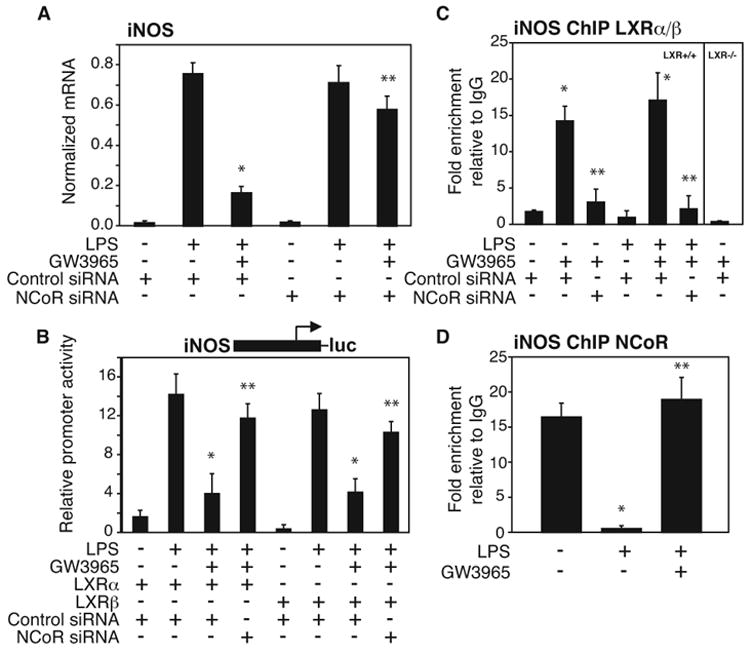
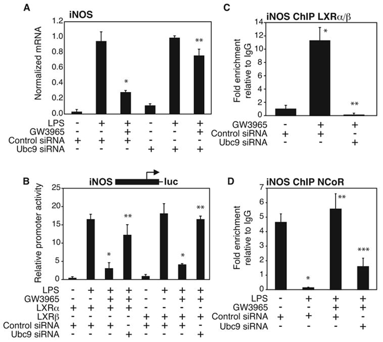
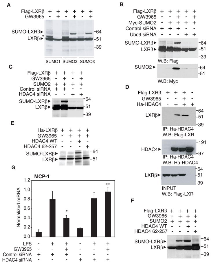
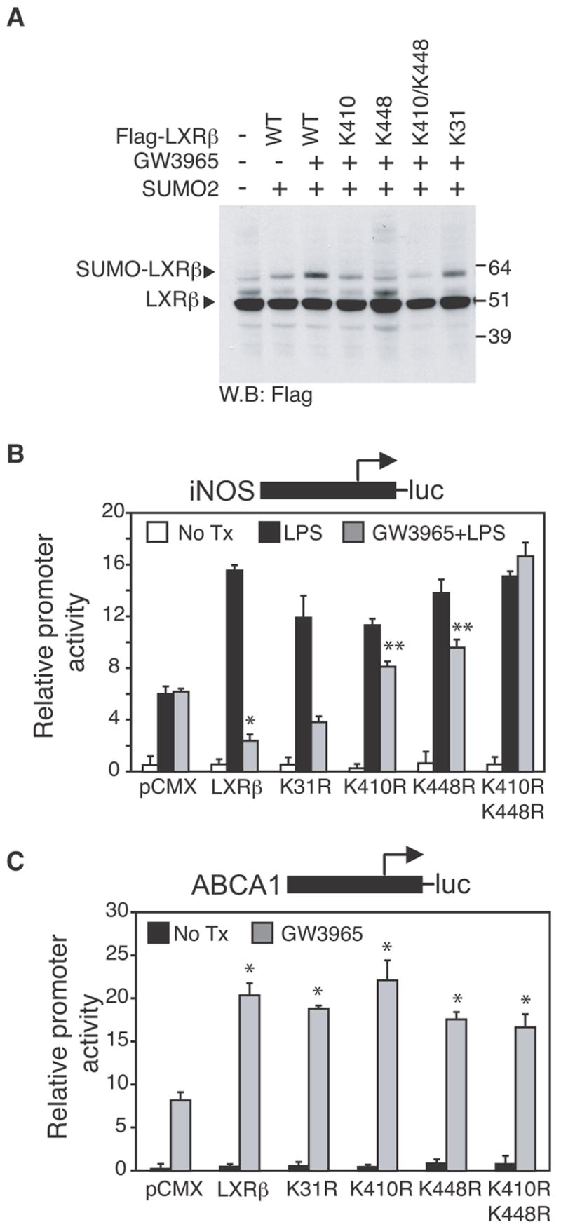
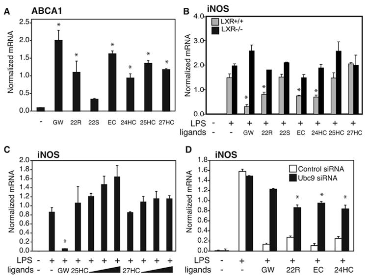
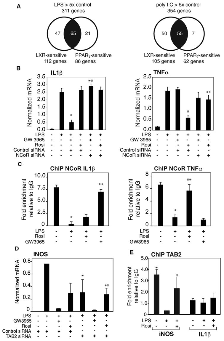
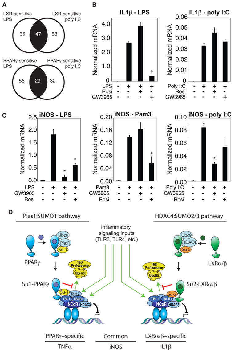
Transrepression is widely utilized to negatively regulate gene expression, but the mechanisms by which different nuclear receptors effect gene- and signal-specific transrepression programs remain poorly understood. Here, we report the identification of alternative SUMOylation-dependent mechanisms that enable PPARgamma and LXRs to negatively regulate overlapping but distinct subsets of proinflammatory genes. Ligand-dependent conjugation of SUMO2/3 to LXRs or SUMO1 to PPARgamma targets them to promoters of TLR target genes, where they prevent the signal-dependent removal of NCoR corepressor complexes required for transcriptional activation. SUMO1-PPARgamma and SUMO2/3-LXRs inhibit distinct NCoR clearance mechanisms, allowing promoter- and TLR-specific patterns of repression. Mutational analysis and studies of naturally occurring oxysterol ligands indicate that the transactivation and SUMOylation-dependent transrepression activities of LXRs can be independently regulated. These studies define parallel but functionally distinct pathways that are utilized by PPARgamma and LXRs to differentially regulate complex programs of gene expression that control immunity and homeostasis.


Figures

Figure 1
Figure 1. LXR transrepression requires NCoR
NCoR-specific siRNA abolishes LXR-dependent repression of LPS-induced endogenous iNOS gene expression in primary macrophages (A) and iNOS-luciferase reporter gene activity in RAW264.7 cells (B). After siRNA transfection, macrophages were stimulated with LPS (1μg/ml) for 6h in the absence or presence of LXR ligand (GW3965, 1μM). iNOS expression in peritoneal macrophages was evaluated by QPCR and iNOS promoter activation in RAW264.7 cells was evaluated by luciferase assay. *p < 0.03 vs LPS+control siRNA; **p < 0.02 vs GW3965+LPS control siRNA. C. ChIP assay reveals ligand-dependent and NCoR-dependent LXR recruitment to the iNOS promoter. Primary macrophages were treated with LPS (1μg/ml) with or without the LXR ligand (GW3965, 1μM) for 1h. ChIP assay was performed with antibody against LXR (both isoforms, α and α) or control IgG. Immunoprecipitated DNA was analyzed by QPCR using primers specific for the iNOS-promoter. Chromatin isolated from LXR−/−mice was used as a negative control for specificity of the LXR antibody. * p < 0.05 vs untreated or LPS alone; **p < 0.03 vs GW3965 or GW3965+LPS control siRNA. D. LXR ligand inhibits LPS-stimulated release of NCoR from the iNOS promoter as shown by ChIP assay in peritoneal macrophages, treated as described in panel C. * p < 0.02 vs untreated; **p < 0.02 vs LPS. Results are expressed as the average of three independent experiments. Error bars represent standard deviations.
Figure 2
Figure 2. LXR-dependent transrepression is SUMOylation dependent
Ubc9-specific siRNA reverses LXR-dependent repression of LPS-induced iNOS expression in primary macrophages (A) and iNOS promoter activation in RAW264.7 cells (B). After siRNA transfection, macrophages were stimulated with LPS (1μg/ml) for 6h in the absence or presence of LXR ligand (GW3965, 1μM). iNOS expression in peritoneal macrophages was evaluated by QPCR and iNOS promoter activation in RAW264.7 was evaluated by luciferase assay. * p < 0.03 vs LPS control siRNA; **p < 0.02 vs GW3965+LPS control siRNA. C,D. siRNA directed against Ubc9 prevents LXR recruitment (C) and abolishes the ability of the LXR agonist to inhibit the NCoR clearance (D) from the iNOS promoter as shown by ChIP. Primary macrophages were treated with LPS (1μg/ml) with or without the LXR ligand (GW3965, 1μM) for 1h. ChIP assay was performed with antibody against LXR or NCoR or control IgG. Immunoprecipitated DNA was analyzed by real time PCR using primers specific for the iNOS-promoter. (C) * p < 0.01 vs untreated + control siRNA; **p < 0.02 vs GW3965 control siRNA. (D) * p < 0.02 vs untreated + control siRNA; **p < 0.02 vs LPS control siRNA, * **p < 0.01 vs GW3965 + LPS control siRNA. Results are expressed as average of 2 independent experiments. Error bars represent standard deviations.
Figure 3
Figure 3. LXRβ is modified by SUMO-2 and SUMO-3
A. LXRβ is SUMOylated by SUMO-2 and SUMO-3. HeLa cells were transfected with FLAG-LXRβ, Ubc9 and with SUMO-1, SUMO-2 or SUMO-3 expression vector (as indicated) and treated with GW3965 for 1h (1μM). Whole cell lysates were immunoblotted for FLAG-tag. B. Knock down of Ubc9 by specific siRNA blocks the SUMO-2 conjugation of LXRβ. HeLa cells were transfected with siControl, siUbc9 (where indicated), FLAG-LXRβ and MYC-SUMO-2 expression vectors. Whole cell lysated were immunoprecipitated with FLAG-antibody and immunoblotted for FLAG-tag and SUMO2- MYC-tag. C. An siRNA directed against HDAC4 blocks LXRβSUMOylation. HeLa cells were transfected with siControl, siHDAC4 (where indicated), FLAG-LXRβ and SUMO-2 expression vectors and treated with GW3965 for 1h (1μM). Whole cell lysates were immunoblotted for FLAG-tag. D. LXRβ and HDAC4 physically interact. HeLa cells were transfected with FLAG-LXRβand Ha-HDAC4 and treated with GW3965 for 1h as indicated. Whole cell lysate was immunoprecipitated with HA antibody (middle panel) and LXRβ was detected with FLAG antibody (upper panel). In the lower panel the INPUT shows an equal expression of LXRβin each sample. Cells transfected with only LXRβ or HDAC4 (first and last lane) were used as negative control for the IP. E. LXRβ is SUMOylated by HDAC4 in vitro. The in vitro SUMOylation assay was performed as described in Experimental Procedures, adding to the reaction HDAC4 or HDAC4 mutant (67-257) as indicated. The proteins were immunoblotted for HA-tag. F. HDAC4 modulates LXRβ SUMOylation in vivo. HeLa cells were transfected with FLAG-LXRβ, Ubc9, SUMO-2 and HDAC4 or HDAC4 mutant (67-257) expression vector (as indicated) and treated with GW3965 for 1h (1μM). Whole cell lysates were immunoblotted for FLAG-tag. G. Knock down of HDAC4 blocks LXR transrepression of MCP1. Primary macrophages were transfected with control or HDAC4 specific siRNA for 48h and then stimulated with LPS (1μg/ml) for 6h in the absence or presence of LXR ligand (GW3965, 1μM). MCP1 expression was evaluated by real time PCR. Results are expressed as average of two independent experiments. Error bars represent standard deviations. * p < 0.02 vs LPS+control siRNA; **p < 0.03 vs GW3965+LPS+ control siRNA.
Figure 4
Figure 4. Mutation of LXRβ SUMO acceptor sites abolishes transrepression but not transactivation activity
A. SUMOylation of LXRβ occurs at K410 and K448, but not at K31, as demonstrated by SUMOylation assay (performed as described in Fig 3A) in HeLa cells transfected with WT FLAG-LXRβ or FLAG-mutants K31, K410, K448 or double mutants K410/K448 as indicated. B,C. LXRβ K410 and K448 are required for transrepression but not for transactivation. RAW264.7 cells were transfected with iNOS-luc (B) or ABCA1-luc (C) reporter plasmids and with an expression vector for LXRβ wild type or mutated in K31, K410, K448 or K410/K448 as indicated. 24h after transfection, cells were treated with DMSO (white bars) or with LPS (1μg/ml) for 12h in the absence (black bars) or presence of LXR ligand (GW3965, 1μM, grey bars). Results are expressed as average of three independent experiments. Error bars represent standard deviations. (B) * p < 0.01 vs LPS; **p < 0.05 vs GW3965+LPS+ LXRβ wild type. (C) * p < 0.03 vs pCMX.
Figure 5
Figure 5. Endogenous LXRs ligands promote entry into transrepression pathway
A,B. Natural LXRs ligands induce ABCA1 expression and repress iNOS activation. Primary macrophages from WT (panel A and B, grey bars) and LXR−/− mice (panel B, black bars) were treated with GW3965 (1μM) or 5μM oxysterols 22R, 22S, 24(S),25EC, 24HC, 25HC and 27HC (A) for 18 hours. In the B panel cells were pretreated with the indicated ligands and then treated with LPS (1μg/ml) for 6h. Expression of ABCA1 (A) and iNOS (B) was evaluated by QPCR. (A) * p < 0.03 vs untreated; (B) * p < 0.01 vs LPS. C. Dose-response experiment of 25HC and 27HC. Primary macrophages were pretreated with GW3965(1μM), 25HC and 27HC at increasing concentrations (1μM, 5 μM, 10 μM, 50 μM) and then treated with LPS (1μg/ml) for 6h. Expression of iNOS was evaluated by QPCR. * p < 0.01 vs LPS. D. Natural LXRs ligands require Ubc9 to transrepress iNOS expression. Primary macrophages were transfected with Ubc9 siRNA (black bars) or non specific siRNA (white bars) for 48h and then stimulated with LPS (1μg/ml) for 6h in the absence or presence of LXR ligands as indicated (5μM). iNOS expression in peritoneal macrophages was evaluated by QPCR. * p < 0.05 vs control siRNA. Results are expressed as average of two independent experiments. Error bars represent standard deviations.
Figure 6
Figure 6. Gene-specific utilization of NCoR and SUMOylation-dependent pathways in LXRs and PPARγ transrepression
A. Relationship of genes subject to transrepression by LXR or PPARγ agonists in primary macrophages stimulated with either LPS or poly I:C as determined by expression profiling assays. Genes were filtered based on at least 5-fold induction by LPS and poly I:C, and at least 50% repression by each PPARγ or LXR agonist. Data is derived from (Ogawa et al., 2005). B. Receptor-specific repression of IL1β and TNFα. Primary macrophages were transfected with siRNA control or NCoR specific siRNA for 48h and then stimulated with LPS (1μg/ml) for 6h in the absence or presence of LXR ligand (GW3965, 1γM) or PPARγ ligand (Rosiglitazone, 1μM). IL1β and TNFα expression was evaluated by QPCR. * p < 0.01 vs LPS; ** p < 0.01 vs GW3965 (left panel) or Rosi (right panel) +LPS control siRNA. C. Differential inhibition of NCoR clearance from IL1β and TNFα promoters by PPARγ and LXR ligands. Primary macrophages were treated with LPS (1μg/ml) with or without PPARγ (Rosiglitazone, 1μM) or LXR ligand (GW3965, 1μM) for 1h. ChIP assays were performed with antibody against NCoR and or control IgG. * p < 0.01 vs untreated; ** p < 0.08 vs LPS. D. PPARγ transrepression is TAB2-dependent. Primary macrophages were transfected with siRNA control or TAB2 specific siRNA for 48h and then stimulated with LPS (1μg/ml) for 6h in the absence or presence of LXR ligand (GW3965, 1μM) or PPARγ ligand (Rosiglitazone, 1μM). iNOS expression was evaluated by QPCR. * p < 0.01 vs LPS; ** p < 0.01 vs Rosi +LPS control siRNA. E. Tab2 is present in the basal state on the iNOS promoter, but not on the IL1β promoter. ChIP assays were performed with antibody against Tab2 or control IgG. * p<0.01 vs control IgG. Results are expressed as the average of 2 independent experiments. Errors bars represent standard deviations.
Figure 7
Figure 7. Signal-specific regulation of NCoR and SUMOylation-dependent transrepression
A. Signal-dependence of repression by PPARγ and LXR agonists. Genes in the central sector of each diagram were repressed when induced by either LPS or poly I:C. Genes in the left sector were only repressed when induced by LPS, while genes in the right sector were only repressed when induced by poly I:C. B. The IL-1β gene is induced by both LPS and poly I:C, but is only sensitive to LXR repression when induced by LPS. Primary macrophages were treated with TLR4 specific agonist (LPS,1μg/ml), or TLR3 specific agonist (polyI:C, 50 ng/ml) for 6h with or without PPARγ (Rosiglitazone, 1μM) or LXR ligand (GW3965, 1μM). IL-1β expression was evaluated by QPCR. * p < 0.02 vs LPS. C. Differential transrepression of the iNOS gene by LXR and PPARγ ligands. Primary macrophages were treated with the LPS (1 μg/ml), TLR2-specific agonist (PAM3, 300 ng/ml) or poly I:C (50 ng/ml) for 6h with or without PPARγ (Rosiglitazone, 1μM) or LXR ligand (GW3965, 1μM). iNOS expression was evaluated by real time PCR. * p < 0.02 vs LPS (left), Pam 3 (middle), poly I:C (right). Results are expressed as the average of 2 independent experiments performed in duplicate. Error bars represent standard deviations. D. Model depicting parallel transrepression pathways utilized by PPARγ and LXRs. See text for discussion.

Comment in

References

    1. Baek SH, Ohgi KA, Rose DW, Koo EH, Glass CK, Rosenfeld MG. Exchange of N-CoR corepressor and Tip60 coactivator complexes links gene expression by NF-kappaB and beta-amyloid precursor protein. Cell. 2002;110:55–67. - PubMed
    1. Castrillo A, Joseph SB, Vaidya SA, Haberland M, Fogelman AM, Cheng G, Tontonoz P. Crosstalk between LXR and toll-like receptor signaling mediates bacterial and viral antagonism of cholesterol metabolism. Mol Cell. 2003;12:805–816. - PubMed
    1. Chang HM, Paulson M, Holko M, Rice CM, Williams BR, Marie I, Levy DE. Induction of interferon-stimulated gene expression and antiviral responses require protein deacetylase activity. Proc Natl Acad Sci U S A. 2004;101:9578–9583. - PMC - PubMed
    1. Chiang JY, Kimmel R, Stroup D. Regulation of cholesterol 7alpha-hydroxylase gene (CYP7A1) transcription by the liver orphan receptor (LXRalpha) Gene. 2001;262:257–265. - PubMed
    1. Costet P, Luo Y, Wang N, Tall AR. Sterol-dependent transactivation of the ABC1 promoter by the liver X Receptor/Retinoid X receptor. J Biol Chem. 2000;275:28240–28245. - PubMed

Publication types

MeSH terms

Associated data