

Differential control of the scapulothoracic muscles in humans
- PMID: 17218352
- PMCID: PMC2075462
- DOI: 10.1113/jphysiol.2006.126276
Differential control of the scapulothoracic muscles in humans
Abstract
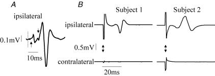
The control of the scapulothoracic muscles trapezius (Tr) and serratus anterior (SA) has been examined in normal human subjects. Electromyographic recordings were made from the SA and Tr muscles (upper trapezius UTr, lower trapezius LTr) using surface electrodes placed bilaterally. Magnetic stimulation of the motor cortex and electrical stimulation of peripheral nerves were used to examine their descending and reflex control. The average optimal site of cortical stimulation was found to be the same for SA, UTr and LTr (an approximate centre of gravity of -0.6 cm, 3.7 cm where the centre of gravity is expressed as the mean anterio-posterior position, the mean medio-lateral position). Some asymmetry in the cortical representation of UTr was found in each individual tested. Magnetic stimulation evoked bilateral MEPs in Tr (latency contralateral (c) UTr 8.5 +/- 1.6 ms, ipsilateral (i) UTr 19.0 +/- 2.7 ms) but only contralateral responses were evoked in SA (11.2 +/- 2.6 ms). Electrical stimulation of the long thoracic nerve at two sites was used to examine homonymous and heteronymous reflexes of SA, while electrical stimulation of cervical nerve of C3/4 was used to examine the heteronymous reflexes of Tr. Ipsilateral SA H reflexes were evoked at a latency of 9.9 +/- 0.8 ms (proximal site) and 10.8 +/- 1.2 ms (distal site). No group I reflexes were evoked from SA to its contralateral homologue. No group I reflexes were evoked between Tr and SA. Finally, cross-correlation of activity from the Tr muscle pairs and the SA muscle pair revealed that the motoneurones of the Tr muscles share some common presynaptic input whereas there was no detectable common presynaptic input to the SA muscle pair. This study extends and consolidates knowledge regarding the neural control of trapezius and for the first time explores the neural control of SA. The study demonstrates a contrasting bilateral control of Tr and SA. These patterns of connections are discussed in relation to the contrasting bilateral functional roles of these muscles.
Figures


References
-
- Alexander CM, Harrison PJ. The bilateral reflex control of the trapezius muscle in humans. Exp Brain Res. 2002;142:418–424. - PubMed
-
- Alexander CM, Harrison PJ. Reflex connections from forearm and hand afferents to shoulder girdle muscles in humans. Exp Brain Res. 2003;148:277–282. - PubMed
-
- Baldissera F, Hultborn H, Illert M. Integration of spinal neuronal systems. In: Brookhart JM, Mountcastle VB, Brooks VB, Geiger SR, editors. Handbook of Physiology section 1, The Nervous System Motor Control. II. Bethesda: American Physiological Society; 1981. pp. 509–595.
-
- Bawa P, Hamm JD, Dhillon P, Gross PA. Bilateral responses of upper limb muscles to transcranial magnetic stimulation in human subjects. Exp Brain Res. 2004;158:385–390. - PubMed
-
- Berardelli A, Priori A, Inghilleri M, Cruccu G, Mercuri B, Manfredi M. Corticobulbar and corticospinal projections to neck muscle motoneurons in man. A functional study with magnetic and electric transcranial brain stimulation. Exp Brain Res. 1991;87:402–406. - PubMed
Publication types
MeSH terms
Grants and funding
LinkOut - more resources
Full Text Sources
Miscellaneous