. 2007 Mar 15;579(Pt 3):691-702.
doi: 10.1113/jphysiol.2006.126094. Epub 2007 Jan 11.

Ionic currents underlying the response of rat dorsal vagal neurones to hypoglycaemia and chemical anoxia

Affiliations

Ionic currents underlying the response of rat dorsal vagal neurones to hypoglycaemia and chemical anoxia

Robert H Balfour et al. J Physiol. .

Abstract

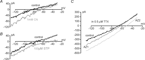
A proportion of dorsal vagal neurones (DVN) are glucosensors. These cells respond to brief hypoglycaemia either with a K(ATP) channel-mediated hyperpolarization or with depolarization owing to an as yet unknown mechanism. K(ATP) currents are observed not only during hypoglycaemia, but also in response to mitochondrial inhibition. Here we show that similarly to the observations for K(ATP) currents, both hypoglycaemia and inhibition of mitochondrial function elicited a small inward current that persisted in TTX in DVN of rat brainstem slices. Removal of glucose from the bath solution induced this inward current within 50 +/- 4 s in one subpopulation of DVN and in 279 +/- 36 s in another subpopulation. No such subpopulations were observed for the response to mitochondrial inhibition. Biophysical analysis revealed that mitochondrial inhibition or hypoglycaemia inhibited an openly rectifying K+ conductance in 25% of DVN. In the remaining cells, either an increase in conductance, with a reversal potential between -58 and +10 mV, or a parallel inward shift of the holding current was observed. This current most probably resulted from inhibition of the Na+-K+-ATPase and/or the opening of an ion channel. Recordings with electrodes containing 145 mm instead of 5 mm Cl- failed to shift the reversal potential of the inward current, indicating that a Cl- channel was not involved. In summary, glucosensing and non-glucosensing DVN appear to use common electrical pathways to respond to mitochondrial inhibition and to hypoglycaemia. We suggest that differences in glucose metabolism rather than differences in the complement of ion channels distinguish these two cell types.


Figures

Figure 2
Figure 2
Inward current elicited by hypoglycaemia or chemical anoxia A, voltage-clamp recording of a GE neurone. Superfusion with glucose-free ACSF (indicated by the bar) led to an inward current (onset indicated by arrow) prior to the development of a KATP channel-mediated outward current. B, a similar inward current prior to KATP current activation was also observed in response to mitochondrial inhibition by 3 mm azide. Plotted is the membrane current at −60 mV every 20 s (black circles). C, an inward current at −60 mV was also elicited by prolonged hypoglycaemia in a slow responding neurone. Plotted is the current at −60 mV during a ramp voltage-clamp protocol. Ramps were applied every 60 s. D, current–voltage relationships from the recording in C at the points indicated. Exposure to glucose-free solution elicited first an inward current with a reversal potential of −30 mV (ii), followed by the KATP current (iii). Note the difference in apparent reversal potential of KATP current when plotted against ii rather than i (arrows).
Figure 1
Figure 1
Transient depolarization of DVN during metabolic inhibition Aa, current-clamp recording showing that application of 3 mm azide (AZ), as indicated by the bar, elicited an initial depolarization and increase in firing rate in a DVN (ii), followed by KATP channel-mediated hyperpolarization and cessation of firing (iii). Ab, current-clamp recording from Aa at a higher time resolution showing: membrane potential (Em) and firing rate in control conditions (i); initial azide-induced depolarization (ii); KATP channel-mediated hyperpolarization and cessation of firing (iii); and recovery (iv). Ba, current-clamp recording from a GE cell showing a small initial depolarization and increase in firing prior to KATP channel opening during superfusion with glucose-free ACSF. Bb, sections of the recording shown in Ba at the points indicated, shown at higher time resolution.
Figure 4
Figure 4
Metabolic inhibition induced inward currents in DVN without functional KATP channels A, voltage-clamp recording of a DVN in which application of 3 mm azide (AZ), as indicated by the bar, elicited a reversible inward current. An outward current was not observed, demonstrating a lack of functional KATP channels. Hypoglycaemia also elicited inward currents in DVN (B and C). B is a plot of Im at −80 mV with readings taken every 5 s. Glucose was removed for a total of 16 min. The inward current started after approximately 4 min as indicated by the open arrow. The inward current was completely reversible after reintroduction of glucose. C, left panel, another DVN exhibited an inward current within < 1 min (open arrow) of exposure to glucose-free ACSF. Right panel, I–V relationships obtained at points i and ii of the recording in the left panel demonstrated that the inward current induced by hypoglycaemia had a reversal potential of approximately −30 mV. D, I–V relationships for a neurone responding fast to hypoglycaemia in the presence of TTX. Inset shows whole-cell current elicited by a 30 ms depolarizing voltage step from −60 to −30 mV in the absence and presence of 0.5 μm TTX. E, there was no significant difference in the time of onset (expressed as mean ±s.e.m.) between the inward currents induced by 1 mm cyanide (CN), 3 mm azide (AZ) and hypoglycaemia for fast responding cells; however, the time of onset for hypoglycaemia in slow responding cells was significantly greater. **P < 0.01. This was the case in both the absence and presence of 0.5 μm TTX. Numbers of recordings are given above bars.
Figure 3
Figure 3
The inward current elicited by metabolic inhibition is independent of KATP currents A, voltage-clamp recording showing an inward current in response to 1 mm cyanide (CN) in a neurone with KATP channels opened by 0.2 mm diazoxide (DZ). Plotted is the mean membrane current at the holding potential of −30 mV every 60 s. B, I–V relationships taken from A (points i, ii and iii), demonstrating that the closure of a K+ current underlies the inward current seen in A. C, I–V relationships showing a cyanide-induced inward current in the presence of 0.1 mm tolbutamide (TB) in a different DVN. Here the inward current reverses at approximately −30 mV.
Figure 5
Figure 5
Biophysical properties of inward currents elicited by metabolic inhibition A, the reversal potential of the inward current elicited by cyanide (CN), azide (AZ) and hypoglycaemia (0 gluc; separate columns for slow and fast responding cells) varied widely between individual neurones. In a subpopulation of DVNs, a decrease in conductance with a reversal potential close to EK was observed during metabolic inhibition (^). Other cells developed an increase in conductance with a wide range of reversal potentials (•). Reversal potentials obtained in the presence of 0.5 μm TTX are given in separate columns indicated by arrows. Cells that exhibited a parallel, depolarizing shift are not represented. Ba, example of a DVN in which 1 mm cyanide inhibited a current with a reversal potential close to EK. Bb, I–V relationships of the current inhibited by cyanide were well fitted by the GHK equation (dashed line). Ca, I–V relationships, in the presence and absence of 1 mm halothane, from a cell in which 1 mm cyanide inhibited a K+ current. Cb, the halothane-sensitive current (difference between I–V relationships shown in Ca) is well described by the GHK equation (dashed line).
Figure 6
Figure 6
Inhibition of the Na+–K+-ATPase A, I–V relationships from a DVN exposed to 1 mm cyanide (CN). B, I–V relationships from the same cell exposed to 100 μm strophanthidin (STP). C, consecutive I–V relationships from a cell exposed to 3 mm azide (AZ), obtained before (control) and after 1 min (AZ1) and 1.5 min of azide exposure (AZ2). Azide first caused an increase in conductance with a reversal potential of −45 mV and then an inward current without apparent reversal potential.

Similar articles

Cited by

References

    1. Ashcroft FM. Adenosine 5′-triphosphate-sensitive potassium channels. Annu Rev Neurosci. 1988;11:97–118. - PubMed
    1. Ashford ML, Boden PR, Treherne JM. Glucose-induced excitation of hypothalamic neurones is mediated by ATP-sensitive K+ channels. Pflugers Arch. 1990;415:479–483. - PubMed
    1. Balfour RH, Hansen AM, Trapp S. Neuronal responses to transient hypoglycaemia in the dorsal vagal complex of the rat brainstem. J Physiol. 2006;570:469–484. - PMC - PubMed
    1. Ballanyi K, Doutheil J, Brockhaus J. Membrane potentials and microenvironment of rat dorsal vagal cells in vitro during energy depletion. J Physiol. 1996;495:769–784. - PMC - PubMed
    1. Ballanyi K, Kulik A. Intracellular Ca2+ during metabolic activation of KATP channels in spontaneously active dorsal vagal neurons in medullary slices. Eur J Neurosci. 1998;10:2574–2585. - PubMed

Publication types

MeSH terms