. 2007 Apr 1;92(7):2583-96.
doi: 10.1529/biophysj.106.094110. Epub 2007 Jan 11.

Upregulation of forces and morphogenic asymmetries in dorsal closure during Drosophila development

Affiliations

Upregulation of forces and morphogenic asymmetries in dorsal closure during Drosophila development

X G Peralta et al. Biophys J. .

Abstract

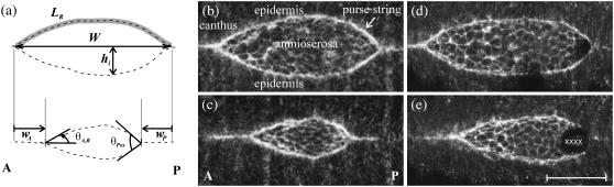
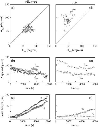
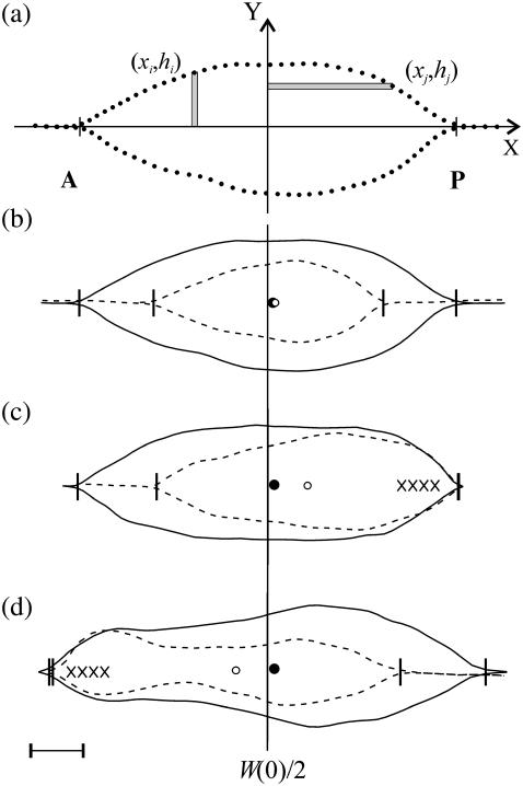
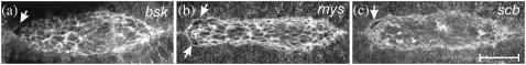
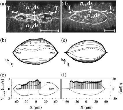
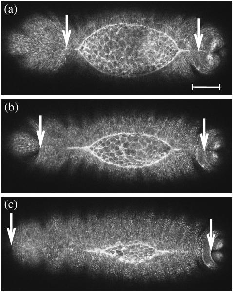
Tissue dynamics during dorsal closure, a stage of Drosophila development, provide a model system for cell sheet morphogenesis and wound healing. Dorsal closure is characterized by complex cell sheet movements, driven by multiple tissue specific forces, which are coordinated in space, synchronized in time, and resilient to UV-laser perturbations. The mechanisms responsible for these attributes are not fully understood. We measured spatial, kinematic, and dynamic antero-posterior asymmetries to biophysically characterize both resiliency to laser perturbations and failure of closure in mutant embryos and compared them to natural asymmetries in unperturbed, wild-type closure. We quantified and mathematically modeled two processes that are upregulated to provide resiliency--contractility of the amnioserosa and formation of a seam between advancing epidermal sheets, i.e., zipping. Both processes are spatially removed from the laser-targeted site, indicating they are not a local response to laser-induced wounding and suggesting mechanosensitive and/or chemosensitive mechanisms for upregulation. In mutant embryos, tissue junctions initially fail at the anterior end indicating inhomogeneous mechanical stresses attributable to head involution, another developmental process that occurs concomitant with the end stages of closure. Asymmetries in these mutants are reversed compared to wild-type, and inhomogeneous stresses may cause asymmetries in wild-type closure.


Figures

FIGURE 1
FIGURE 1
(a) Geometric parameters that describe dorsal closure, where dashed lines are contours of the leading edges (see text). The arc length LR is indicated by the shaded line. (b,c) Confocal fluorescent images of native closure taken 1700 s apart. (d,e) Images of the single-canthus nicking protocol targeted to the posterior end, also taken 1700 s apart. In frame d, dark patches are evident near the left purse-string and the posterior canthus. Dark patches were observed occasionally and are due to the dorsal surface lying just outside the focal plane; however, scanning the focal plane verified the tissues were continuous. In frame e, the X characters indicate the location and extent of the laser perturbations. Scale bar is 50 μm. The anterior end (A) is to the left in all figures.
FIGURE 2
FIGURE 2
Geometric parameters of the dorsal opening for (ac) a wild-type, unperturbed embryo, and (df) a scab mutant (see text). In panels a and d, each circle represents a time point. Solid lines in panel c are the result of the fit based on Eq. 5 and detailed in Appendix B. A fit was not possible in panel f due to complications in implementing Eqs. B-17–B-19 in the analysis; specifically, the data set was too sparse for numerical convergence.
FIGURE 3
FIGURE 3
(a) Contour of the leading edges during native closure showing examples of hiΔx and xjΔy used in the calculation of the centroid xc. Points are the result of the active contour algorithm and indicate the stepping procedure (see text). (b) Contours of the leading edges during native, unperturbed closure, where the dashed contours were taken 1700 s after the initial solid contours. Circles indicate xc at the earlier (solid) or later (open) times. (c) Contours of the single-canthus nicking protocol targeted to the posterior canthus, where the initial contour is just before the first laser incision. (d) Contours of the single-canthus nicking protocol targeted to the anterior canthus, where the initial contour is ∼900 s after the first laser incision. The vertical line indicates the location of W(0)/2 and the vertical ticks locate the canthi. Scale bar is 20 μm.
FIGURE 4
FIGURE 4
Dorsal closure in (a) bsk, (b) mys, and (c) scb mutant embryos shown at the onset of failure. The arrows locate the site of failure. Scale bar is 50 μm.
FIGURE 5
FIGURE 5
Determination of vrecoil in the presence of a localized constriction due to the double-canthi nicking protocol (ac) and in native, unperturbed closure (df). In panels a and d, confocal fluorescent images with free body diagrams superimposed. Scale bar is 50 μm. In panels b and e, digitized contours showing the recoil of the purse-strings after an edge-cut protocol (dashed lines), where the innermost contour is just before the edge-cut and subsequent contours progress from inside to outside in 10.8 s steps. The total time is 140 s. In panels c and f, vrecoil after edge-cuts. Vertical arrows represent vrecoil and correspond to the far-left axis. Bold lines indicate the contour of the purse-strings just before the edge-cut where height corresponds to the far-right axis. The X characters in panels a–c indicate the location and extent of the laser perturbations. In panel c, the dotted lines indicate the boundary of the intact amnioserosa.
FIGURE 6
FIGURE 6
Confocal fluorescent images of native closure showing head involution. The arrows track the movement of anterior and posterior epidermal tissue boundaries. Images were taken 46 min apart. Scale bar is 50 μm.
FIGURE 7
FIGURE 7
Schematic representation of the setting sun model. Two intersecting circular arcs representing native dorsal closure at earlier (left) and later (right) stages. Shown in bold are the arclength L/2; the half-horizontal opening W/2; the half-vertical opening at the symmetry point h; the half-opening angle θ; and the radius r.
FIGURE 8
FIGURE 8
Time dependence of various parameters that describe changes in the geometry of the dorsal opening. The solid (open) circles are for the right (left) leading edge. (a) Curvature (κ) of the leading edge. (Inset) fitted arcs at 0, 3000, and 5000 s, where the extent of the upper arc corresponds to the length of the leading edge at 0 s. The arc at 3000 s essentially is superposed on the arc at 0 s. Tick marks correspond to the lengths of the leading edge at 3000 s (upper arc) and 5000 s (lower arc), respectively. (b) Arclengths of the leading edges L and canthus-to-canthus distance W (crosses). (c) Ratio of 1+RW to 1+Rh. (d) dL/dt for the right (solid line) and left (dashed line) leading edges.
FIGURE 9
FIGURE 9
Determination of the recoil velocity. (a,b) Analysis of the recoil in h(x, t) after the edge-cut protocols. Insets highlight the determination of teff at the intersection of the two fits. Plots of h(x, t) are at (a) x = 6 μm in Fig. 5 c, and (b) x = 28 μm in Fig. 5 f.

References

    1. Keller, R., L. A. Davidson, and D. R. Shook. 2003. How we are shaped: the biomechanics of gastrulation. Differentiation. 71:171–205. - PubMed
    1. Kiehart, D. P., C. G. Galbraith, K. A. Edwards, W. L. Rickoll, and R. A. Montague. 2000. Multiple forces contribute to cell sheet morphogenesis for dorsal closure in Drosophila. J. Cell Biol. 149:471–490. - PMC - PubMed
    1. Jacinto, A., W. Wood, T. Balayo, M. Turmaine, A. Martinez-Arias, and P. Martin. 2000. Dynamic actin-based epithelial adhesion and cell matching during Drosophila dorsal closure. Curr Biol. 10:1420–1426. - PubMed
    1. Hutson, M. S., Y. Tokutake, M.-S. Chang, J. W. Bloor, S. Venakides, D. P. Kiehart, and G. S. Edwards. 2003. Forces for morphogenesis investigated with laser microsurgery and quantitative modeling. Science. 300:145–149. - PubMed
    1. Jacinto, A., S. Woolner, and P. Martin. 2002. Dynamic analysis of dorsal closure in Drosophila: from genetics to cell biology. Dev. Cell. 3:9–19. - PubMed

Publication types

LinkOut - more resources