

Hypochlorite-modified albumin colocalizes with RAGE in the artery wall and promotes MCP-1 expression via the RAGE-Erk1/2 MAP-kinase pathway
- PMID: 17218539
- PMCID: PMC4864469
- DOI: 10.1096/fj.06-7439com
Hypochlorite-modified albumin colocalizes with RAGE in the artery wall and promotes MCP-1 expression via the RAGE-Erk1/2 MAP-kinase pathway
Abstract
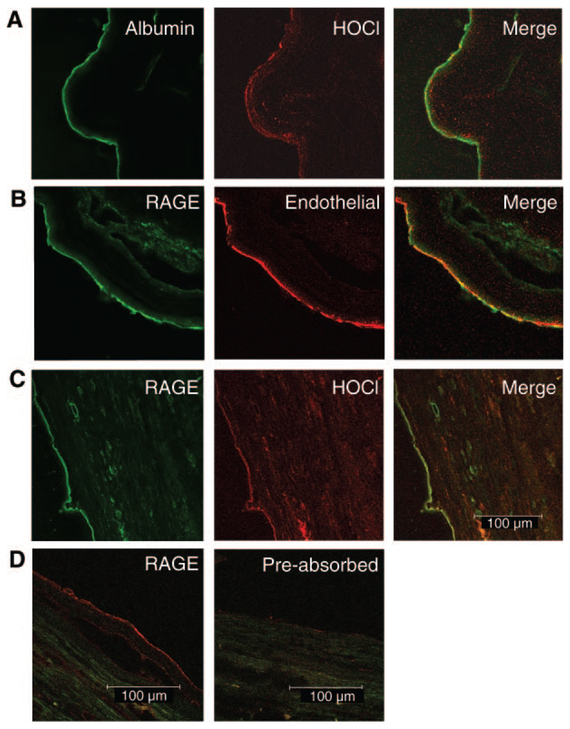
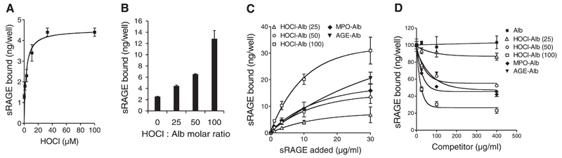
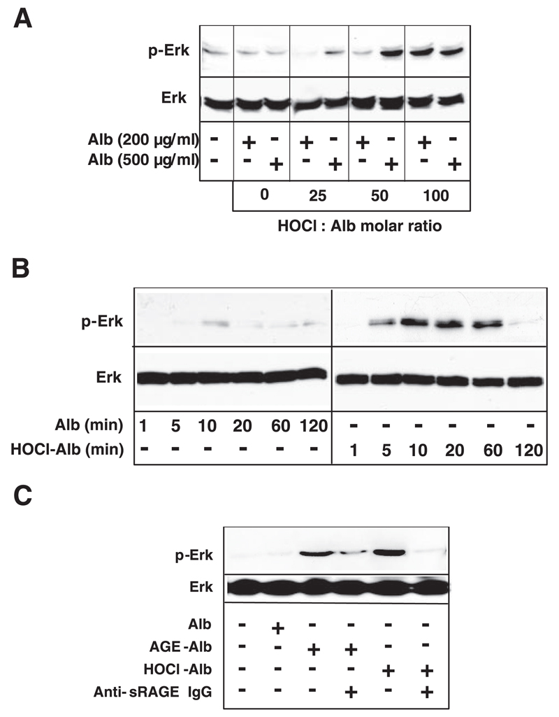
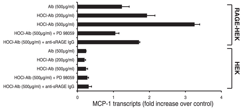
Signal transduction via the endothelial receptor for advanced glycation end products (RAGE) plays a key role in vascular inflammation. Recent observations have shown that the myeloperoxidase-H2O2-chloride system of activated phagocytes is highly up-regulated under inflammatory conditions where hypochlorous acid (HOCl) is formed as the major oxidant. Albumin, an in vivo carrier for myeloperoxidase is highly vulnerable to oxidation and a major representative of circulating advanced oxidized proteins during inflammatory diseases. Immunohistochemical studies performed in the present study revealed marked colocalization of HOCl-modified epitopes with RAGE and albumin in sections of human atheroma, mainly at the endothelial lining. We show that albumin modified with physiologically relevant concentrations of HOCl, added as reagent or generated by the myeloperoxidase-H2O2-chloride system, is a high affinity ligand for RAGE. Albumin, modified by HOCl in the absence of free amino acids/carbohydrates/lipids to exclude formation of AGE-like structures, induced a rapid, RAGE-dependent activation of extracellular signal-regulated kinase 1/2 and up-regulation of the proinflammatory mediator monocyte chemoattractant protein-1. Cellular activation could be blocked either by a specific polyclonal anti-RAGE IgG and/or a specific mitogen-activated protein-kinase kinase inhibitor. The present study demonstrates that HOCl-modified albumin acts as a ligand for RAGE and promotes RAGE-mediated inflammatory complications.
Figures
Similar articles
-
Soluble RAGE blocks scavenger receptor CD36-mediated uptake of hypochlorite-modified low-density lipoprotein.FASEB J. 2007 Oct;21(12):3075-82. doi: 10.1096/fj.07-8316com. Epub 2007 May 29. FASEB J. 2007. PMID: 17536039 Free PMC article.
-
Requirement for p38 and p44/p42 mitogen-activated protein kinases in RAGE-mediated nuclear factor-kappaB transcriptional activation and cytokine secretion.Diabetes. 2001 Jun;50(6):1495-504. doi: 10.2337/diabetes.50.6.1495. Diabetes. 2001. PMID: 11375353
-
Overexpression of receptor for advanced glycation end products induces monocyte chemoattractant protein-1 expression in rat vascular smooth muscle cell line.J Atheroscler Thromb. 2012;19(1):13-22. doi: 10.5551/jat.9472. Epub 2011 Nov 12. J Atheroscler Thromb. 2012. PMID: 22082983
-
The receptor for advanced glycation end-products has a central role in mediating the effects of advanced glycation end-products on the development of vascular disease in diabetes mellitus.Nephrol Dial Transplant. 1996;11 Suppl 5:13-6. doi: 10.1093/ndt/11.supp5.13. Nephrol Dial Transplant. 1996. PMID: 9044300 Review.
-
The reactions of hypochlorous acid, the reactive oxygen species produced by myeloperoxidase, with lipids.Acta Biochim Pol. 2000;47(4):889-99. Acta Biochim Pol. 2000. PMID: 11996112 Review.
Cited by
-
[Association of plasma level of advanced oxidation protein products with Framingham risk score in type 2 diabetic patients without vascular diseases].Nan Fang Yi Ke Da Xue Xue Bao. 2018 May 20;38(5):620-624. doi: 10.3969/j.issn.1673-4254.2018.05.19. Nan Fang Yi Ke Da Xue Xue Bao. 2018. PMID: 29891462 Free PMC article. Chinese.
-
Oxidized plasma albumin promotes platelet-endothelial crosstalk and endothelial tissue factor expression.Sci Rep. 2016 Feb 24;6:22104. doi: 10.1038/srep22104. Sci Rep. 2016. PMID: 26905525 Free PMC article.
-
Plasma-advanced oxidation protein products are potent high-density lipoprotein receptor antagonists in vivo.Circ Res. 2009 Mar 27;104(6):750-7. doi: 10.1161/CIRCRESAHA.108.193169. Epub 2009 Jan 29. Circ Res. 2009. PMID: 19179658 Free PMC article.
-
Chlorine gas exposure causes systemic endothelial dysfunction by inhibiting endothelial nitric oxide synthase-dependent signaling.Am J Respir Cell Mol Biol. 2011 Aug;45(2):419-25. doi: 10.1165/rcmb.2010-0151OC. Epub 2010 Dec 3. Am J Respir Cell Mol Biol. 2011. PMID: 21131444 Free PMC article.
-
Cyanate is a novel inducer of endothelial icam-1 expression.Antioxid Redox Signal. 2012 Jan 15;16(2):129-37. doi: 10.1089/ars.2011.4090. Epub 2011 Oct 14. Antioxid Redox Signal. 2012. PMID: 21838543 Free PMC article.
References
-
- Wautier JL, Schmidt AM. Protein glycation. A firm link to endothelial cell dysfunction. Circ Res. 2004;95:233–238. - PubMed
-
- Witko-Sarsat V, Friedlander M, Capeillere-Blandin C, Nguyen-Khoa T, Nguyen AT, Zingraff J, Jungers P, Descamps-Latscha B. Advanced oxidation protein products as a novel marker of oxidative stress in uremia. Kidney Int. 1996;49:1304–1313. - PubMed
-
- Witko-Sarsat V, Friedlander M, Nguyen Khoa T, Capeillere-Blandin C, Nguyen AT, Canteloup S, Dayer JM, Jungers P, Drueke T, Descamps-Latscha B. Advanced oxidation protein products as novel mediators of inflammation and monocyte activation in chronic renal failure. J Immunol. 1998;161:2524–2532. - PubMed
-
- Capeillere-Blandin C, Gausson V, Descamps-Latscha B, Witko-Sarsat V. Biochemical and spectrophotometric significance of advanced oxidized protein products. Biochim Biophys Acta. 2004;1689:91–102. - PubMed
Publication types
MeSH terms
Substances
Grants and funding
LinkOut - more resources
Full Text Sources
Research Materials
Miscellaneous
