. 2007 Apr;292(4):H1664-74.
doi: 10.1152/ajpheart.01138.2006. Epub 2007 Jan 12.

NADPH oxidase modulates myocardial Akt, ERK1/2 activation, and angiogenesis after hypoxia-reoxygenation

Affiliations

NADPH oxidase modulates myocardial Akt, ERK1/2 activation, and angiogenesis after hypoxia-reoxygenation

Jian-Xiong Chen et al. Am J Physiol Heart Circ Physiol. 2007 Apr.

Abstract

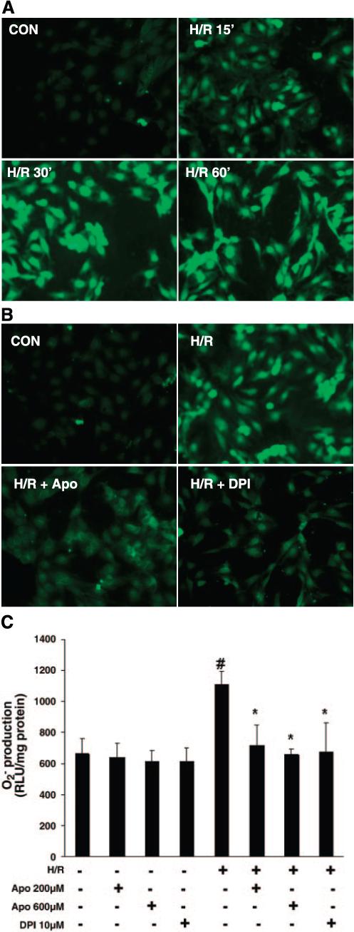
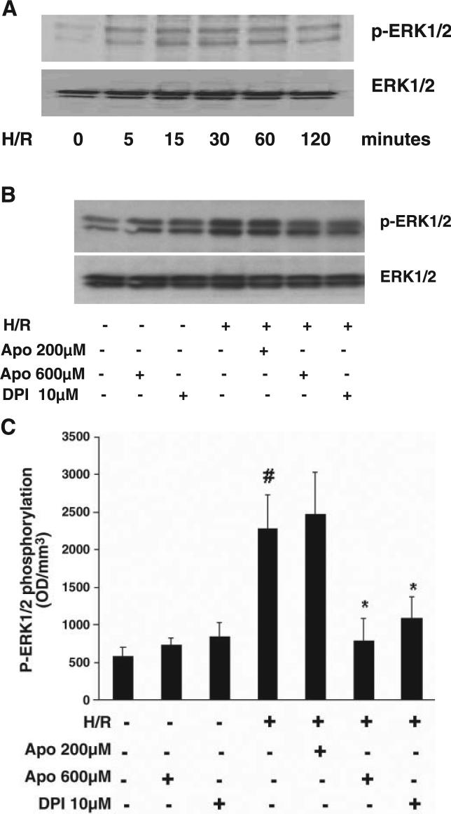
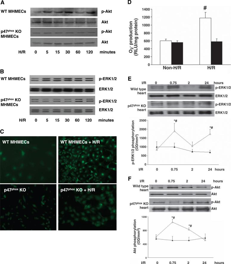
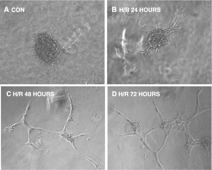
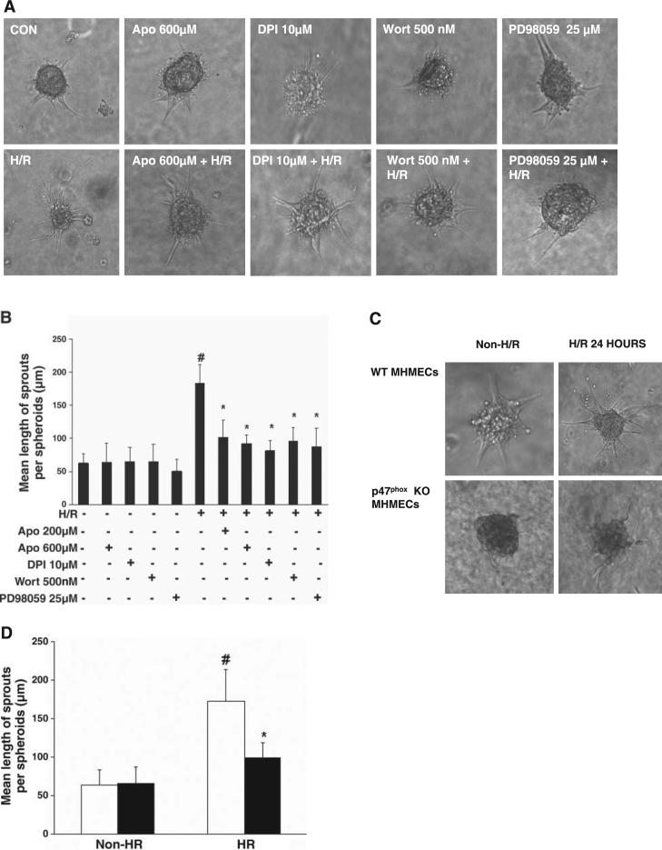
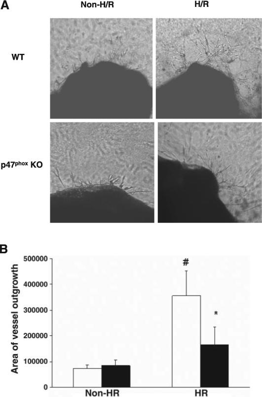
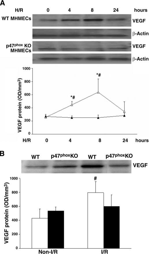
Recent studies have demonstrated that reactive oxygen species (ROS) mediate myocardial ischemia-reperfusion (I/R) and angiogenesis via the mitogen-activated protein kinases and the serine-threonine kinase Akt/protein kinase B pathways. NADPH oxidases are major sources of ROS in endothelial cells and cardiomyocytes. In the present study, we investigated the role of NADPH oxidase-derived ROS in hypoxia-reoxygenation (H/R)-induced Akt and ERK1/2 activation and angiogenesis using porcine coronary artery endothelial cells (PCAECs) and a mouse myocardial I/R model. Our data demonstrate that exposure of PCAECs to hypoxia for 2 h followed by 1 h of reoxygenation significantly increased ROS formation. Pretreatment with the NADPH oxidase inhibitors, diphenyleneiodonium (DPI, 10 microM) and apocynin (Apo, 200 and 600 microM), significantly attenuated H/R-induced ROS formation. Furthermore, exposure of PCAECs to H/R caused a significant increase in Akt and ERK1/2 activation. Exposure of PCAEC spheroids and mouse aortic rings to H/R significantly increased endothelial spheroid sprouting and vessel outgrowth, whereas pharmacological inhibition of NADPH oxidase or genetic deletion of the NADPH oxidase subunit, p47(phox) (p47(phox-/-)), significantly suppressed these changes. With the use of a mouse I/R model, our data further show that the increases in myocardial Akt and ERK1/2 activation and vascular endothelial growth factor (VEGF) expression were markedly blunted in the p47(phox-/-) mouse subjected to myocardial I/R compared with the wild-type mouse. Our findings underscore the important role of NADPH oxidase and its subunit p47(phox) in modulating Akt and ERK1/2 activation, angiogenic growth factor expression, and angiogenesis in myocardium undergoing I/R.


Figures

Fig. 1
Fig. 1
A: time course of hypoxia-reoxygenation (H/R)-stimulated generation of reactive oxygen species (ROS) in porcine coronary artery endothelial cells (PCAECs). Representative images from 3 separate studies of dichlorofluorescein fluorescence in chloromethyl-2′,7′-dichlorodihydrofluorescein diacetate-loaded PCAECs taken at baseline and after exposure to 2 h of hypoxia followed by 15, 30, or 60 min of reoxygenation. B: pretreatment of PCAECs with either diphenyleneiodonium (DPI; 10 μM) or apocynin (Apo; 600 μM) caused an inhibition of H/R-induced ROS generation. C: pretreatment of PCAECs with either the NADPH oxidase inhibitors DPI (10 μM) or Apo (200 and 600 μM), followed by exposure to hypoxia for 2 h and reperfusion for 60 min, resulted in significant inhibition of H/R-induced superoxide formation as measured by lucigenin-dependent chemiluminescence (n = 3−4 cell lines, data are means ± SD. *P < 0.05 compared with H/R; #P < 0.05 compared with baseline). RLU, relative light units; Con, control.
Fig. 2
Fig. 2
A: Western blot analysis showing time course of H/R-stimulated Akt phosphorylation in PCAECs. Phosphorylation of Akt was increased and peaked at 15 and 30 min; by 120 min phospho (p)-Akt had declined toward normal. Total Akt levels were unaltered over 120 min of reoxygenation. B: effect of NADPH oxidase inhibitors DPI (10 μM) or Apo (200 and 600 μM) on H/R-stimulated Akt phosphorylation. C: densitometric data from Western blot analyses of PCAECs showing that pretreatment with DPI and Apo significantly suppressed H/R-induced Akt phosphorylation (n = 3−4 cell lines, data are means ± SD, *P < 0.05 compared with H/R; #P < 0.05 compared with baseline). OD, optical density.
Fig. 3
Fig. 3
A: Western blot analysis showing the time course of H/R-stimulated ERK1/2 phosphorylation in PCAECs. Increased phosphorylation was apparent at 5 min, peaked at 15 min, and remained high over the 120 min of reoxygenation. B: effect of NADPH oxidase inhibitors DPI (10 μM) or Apo (200 and 600 μM) on H/R-stimulated ERK1/2 phosphorylation. Both DPI (10 μM) and Apo (600 μM) but not 200 μM Apo suppressed H/R-stimulated ERK1/2 phosphorylation. C: densitometric data from Western blot analyses of PCAECs showing that pretreatment with DPI (10 μM) and Apo (600 μM), but not Apo (200 μM), significantly suppressed H/R-induced ERK1/2 phosphorylation (n = 4 cell lines, data are means ± SD, *P < 0.05 compared with H/R; #P < 0.05 compared with baseline).
Fig. 4
Fig. 4
Effect of H/R on Akt, ERK1/2 phosphorylation, and ROS formation in wild-type (WT) and p47phox−/− mouse heart microvascular endothelial cells (MHMECs). A: exposure of WT MHMECs to hypoxia for 2 h followed by reoxygenation for 5, 15, 30, 60, and 120 min resulted in a gradual increase in Akt phosphorylation. p47phox−/− MHMECs failed to show an increase in Akt phosphorylation in response to H/R (n = 3 cell lines). B: time course of H/R-stimulated ERK1/2 phosphorylation in WT and p47phox−/− MHMECs. Exposure of WT MHMECs to H/R resulted in a significant increase in phosphorylated ERK1/2. Cells isolated from p47phox−/− mice failed to show an increase in ERK1/2 phosphorylation (n = 3 cell lines). C: H/R- induced intracellular ROS formation was markedly attenuated in p47phox−/− MHMECs compared with WT MHMECs (n = 4 −5 cell lines). D: H/R-induced superoxide production was significantly suppressed in p47phox−/− MHMECs (black bars) as compared with WT MHMECs (white bars) (n = 5 cell lines, data are means ± SD, #P < 0.05 compared with baseline). E and F: time course and patterns of I/R-induced ERK1/2 (E) and Akt (F) phosphorylation in WT (△) and p47phox−/− (▲) mice [n = 4−5 mice, data are means ± SD, *P < 0.05 compared with ischemia-reperfusion (I/R); #P < 0.05 compared with control]. KO, knockout.
Fig. 5
Fig. 5
Representative images showing the sprouting of capillary-like structures from collagen-embedded PCAEC spheroids over time following exposure to H/R. A: 24-h control. B: H/R, 24 h. C: H/R, 48 h. D: H/R, 72 h. Untreated PCAEC spheroids had a low level of spontaneous sprouting. Twenty-four hours of exposure to H/R led to growth of sprouts from the spheroids. Exposure to H/R for 48−72 h resulted in a complex network of tubules sprouting from the spheroids.
Fig. 6
Fig. 6
A: representative images of the effects of inhibitors of NADPH oxidase, phosphatidylinositol 3-kinase (PI3K), and ERK1/2 on H/R-stimulated sprouting from PCAEC spheroids at 24 h. B: quantitative analysis of sprout length from PCAEC spheroids exposed to H/R. Treatment with DPI (10 μM), Apo (200 μm and 600 μM), Wortmannin (Wort; 500nM), or PD-98059 (25 μM) suppressed the H/R-stimulated increase in sprout length (n = 4 cell lines, data are means ± SD; *P < 0.05 compared with H/R; #P < 0.05 compared with baseline). C: images of WT and p47phox−/− MHMEC spheroids exposed to H/R for 24 h. D: quantitative analysis of sprout length from WT (white bars) and p47phox−/− (black bars) MHMEC spheroids exposed to H/R. H/R was less effective in stimulating spheroid spourting in p47phox−/− MHMECs compared with WT MHMECs (n = 3 cell lines, data are means ± SD, *P < 0.05 compared with H/R; #P < 0.05 compared with non-H/R).
Fig. 7
Fig. 7
A: images of the effects of NADPH oxidase inhibitors on H/R-stimulated vessel outgrowth from WT and p47phox−/− mouse aortic rings at days 5−7. WT aortic rings exposed to H/R demonstrated increases in vessel outgrowth at 5−7 days, whereas vessel outgrowth from p47phox−/− mouse aortic rings was minimal at days 5−7. B: quantitative analysis of the area of vessel outgrowth (in arbitrary units) from WT (white bars) and p47phox−/− (black bars) mouse aortic rings. Exposure to H/R significantly increased the outgrowth area in WT mouse aortic rings. The outgrowth area was also increased in the p47phox−/− mouse aortic rings exposed to H/R compared with baseline, but this increase was less than that in H/R-stimulated WT aortic rings (n = 4−5, data are means ± SD, *P < 0.05 compared with H/R; #P < 0.05 compared with baseline).
Fig. 8
Fig. 8
A: Western blot analysis showing time course and patterns of H/R-induced VEGF expression in MHMECs isolated from WT or p47phox−/− mice. Exposure of WT MHMECs (△) to H/R increased VEGF expression at 4 and 8 h, whereas exposure of p47phox−/− MHMECs (▲) to H/R failed to induce VEGF expression (n = 3 cell lines, data are means ± SD, *P < 0.05 compared with H/R; #P < 0.05 compared with baseline). B: Western blot analysis reveals that exposure of I/R increases myocardial VEGF expression in WT (white bars) but not in p47phox−/− mice (black bars, n = 4−5 mice, data are means ± SD, #P < 0.05 compared with control).

References

    1. Abid MR, Kachra Z, Spokes KC, Aird WC. NADPH oxidase activity is required for endothelial cell proliferation and migration. FEBS Lett. 2000;486:252–256. - PubMed
    1. Arbiser JL, Petros J, Klafter R, Govindajaran B, McLaughlin ER, Brown LF, Cohen C, Moses M, Kilroy S, Arnold RS, Lambeth JD. Reactive oxygen generated by Nox1 triggers the angiogenic switch. Proc Natl Acad Sci USA. 2002;99:715–720. - PMC - PubMed
    1. Bell RM, Cave AC, Johar S, Hearse DJ, Shah AM, Shattock MJ. Pivotal role of NOX-2-containing NADPH oxidase in early ischemic preconditioning. FASEB J. 2005;19:2037–2039. - PubMed
    1. Brar BK, Jonassen AK, Stephanou A, Santilli G, Railson J, Knight RA, Yellon DM, Latchman DS. Urocortin protects against ischemic and reperfusion injury via a MAPK-dependent pathway. J Biol Chem. 2000;275:8508–8514. - PubMed
    1. Chen JX, Lawrence ML, Cunningham G, Christman BW, Meyrick B. HSP90 and Akt modulate Ang-1-induced angiogenesis via NO in coronary artery endothelium. J Appl Physiol. 2004;96:612–620. - PubMed

Publication types

MeSH terms

LinkOut - more resources