. 2007 Jan;19(1):226-36.
doi: 10.1105/tpc.106.047613. Epub 2007 Jan 12.

The kinesin ATK5 functions in early spindle assembly in Arabidopsis

Affiliations

The kinesin ATK5 functions in early spindle assembly in Arabidopsis

J Christian Ambrose et al. Plant Cell. 2007 Jan.

Abstract

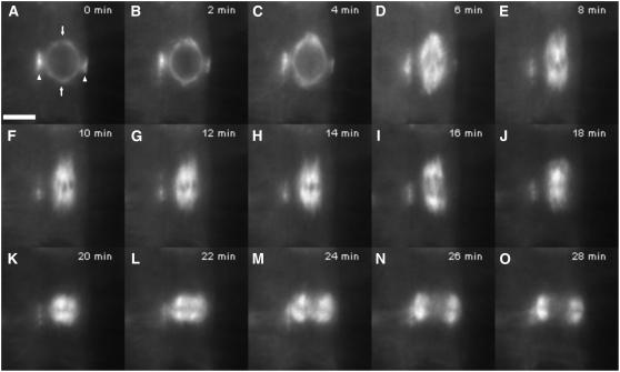
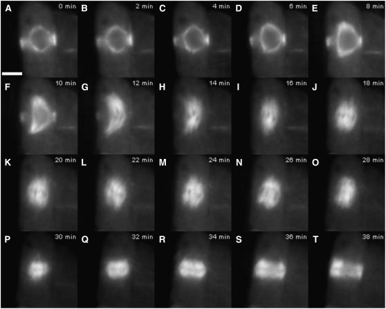
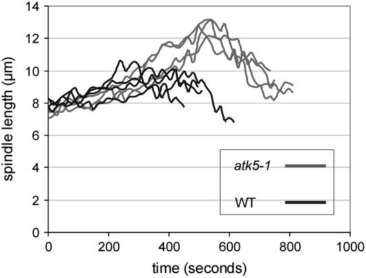
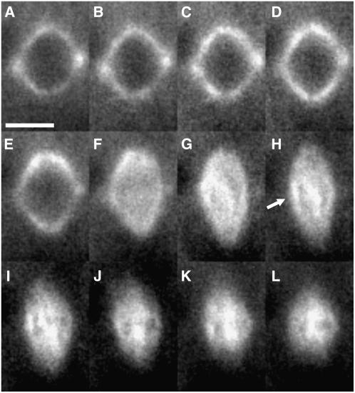
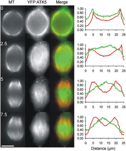
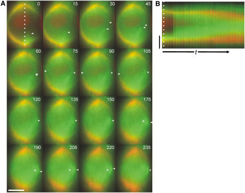
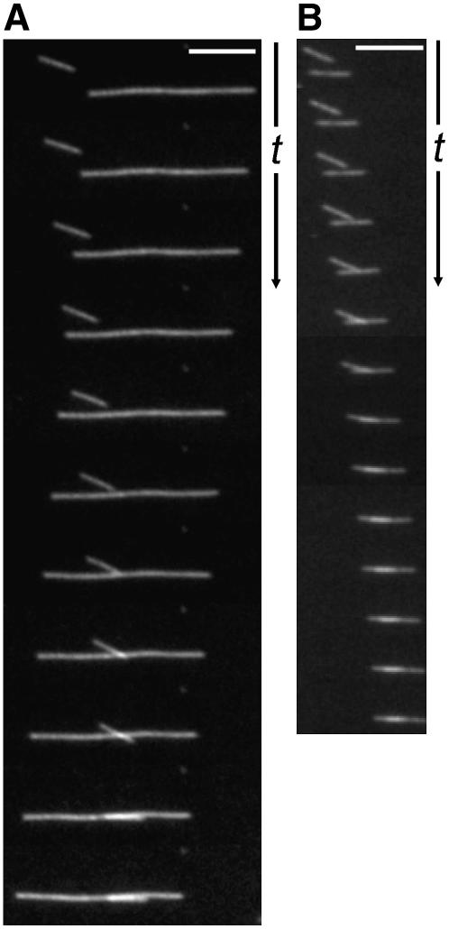
During cell division, the mitotic spindle partitions chromosomes into daughter nuclei. In higher plants, the molecular mechanisms governing spindle assembly and function remain largely unexplored. Here, live cell imaging of mitosis in Arabidopsis thaliana plants lacking a kinesin-14 (ATK5) reveals defects during early spindle formation. Beginning during prophase and lasting until late prometaphase, spindles of atk5-1 plants become abnormally elongated, are frequently bent, and have splayed poles by prometaphase. The period of spindle elongation during prophase and prometaphase is prolonged in atk5-1 cells. Time-lapse imaging of yellow fluorescent protein:ATK5 reveals colocalization with perinuclear microtubules before nuclear envelope breakdown, after which it congresses inward from the poles to the midzone, where it becomes progressively enriched at regions of overlap between antiparallel microtubules. In vitro microtubule motility assays demonstrate that in the presence of ATK5, two microtubules encountering one another at an angle can interact and coalign, forming a linear bundle. These data indicate that ATK5 participates in the search and capture of antiparallel interpolar microtubules, where it aids in generating force to coalign microtubules, thereby affecting spindle length, width, and integrity.


Figures

Figure 1.
Figure 1.
Mitosis in a Wild-Type Arabidopsis Plant Expressing GFP:TUB6. Images are taken from a time-lapse series (see Supplemental Movie 1 online). (A) Prophase spindle with narrow PPB. Arrowheads indicate PPB, and arrows indicate incipient spindle poles. Bar = 5 μm. (B) and (C) Prophase spindle elongation (B) and NEB (C). (D) and (E) Prometaphase. (F) to (H) Metaphase. (I) Anaphase. (J) to (O) Telophase/cytokinesis, showing the phragmoplast expanding centrifugally.
Figure 2.
Figure 2.
Aberrant Mitosis in an atk5-1 Plant Expressing GFP:TUB6. Images are taken from a time-lapse series (see Supplemental Movie 2 online). (A) Prophase spindle with PPB. Bar = 5 μm. (B) to (E) Prophase spindle elongation and NEB. (F) to (I) Prometaphase. (J) to (M) Metaphase. (N) Anaphase. (O) to (T) Telophase/cytokinesis.
Figure 3.
Figure 3.
Spindles of atk5-1 Plants Exhibit an Increased Duration of Prophase/Prometaphase Spindle Elongation. Spindle lengths from four cells of the wild type (black lines) and atk5-1 (gray lines) were measured every 15 s during prophase/prometaphase spindle elongation. The peak of each line represents maximum prometaphase spindle length, after which MTs congress inward to the equator to form the metaphase spindle.
Figure 4.
Figure 4.
Time Series of YFP:ATK5 Localization during Mitotic Spindle Formation in an Arabidopsis Root Tip Cell. (A) and (B) Prophase spindle. YFP:ATK5 localizes to MTs of the prophase spindle and PPB. Bar = 5 μm. (C) to (E) Prophase spindle elongation. (F) NEB. (G) to (I) Prometaphase. (J) to (L) Metaphase. After NEB (F), YFP:ATK5 becomes enriched on interpolar MT bundles traversing the midzone (arrow in [H]). Intervals between frames are 1 min.
Figure 5.
Figure 5.
Localization of YFP:ATK5 (Green) during the Transition from Prophase to Early Prometaphase in a Tobacco BY-2 Cell Expressing MBD:Ds Red (Red) as a MT Reporter. Images are taken from a time-lapse series at 2.5-min intervals (see Supplemental Movie 3 online). Fluorescence intensity profiles for each time point correspond to absolute pixel values for each fluorophore on a line drawn along the long axis of the spindle (MTs = red, YFP:ATK5 = green). At 0 min, YFP:ATK5 colocalizes evenly with MTs of the prophase spindle, which surrounds the nucleus. After NEB, YFP:ATK5 becomes progressively enriched on interpolar MT bundles spanning the midzone while becoming proportionately depleted from the poles relative to MT fluorescence (2.5 to 7.5 min). Bar = 10 μm.
Figure 6.
Figure 6.
Localization of YFP:ATK5 (Green) and MBD:Ds Red (Red) during the Prophase-to-Prometaphase Transition in a Tobacco BY-2 Cell. (A) Images taken from a time-lapse series (see Supplemental Movie 4 online) with 15-s intervals between frames. YFP:ATK5 moves from the poles inward (arrowheads) and becomes enriched at regions of overlap between antiparallel interpolar MT bundles (asterisks). (B) Kymograph of the entire sequence taken along the dotted line in (A). The time scale (t) in (B) is 345 s. Bars = 10 μm.
Figure 7.
Figure 7.
MTs Interact and Coalign in Vitro in the Presence of GST:ATK5 and ATP. (A) Interaction between antiparallel MTs. (B) Interaction between parallel MTs. The time interval between frames is 5 s. Bars = 5 μm.

Similar articles

Cited by

References

    1. Ambrose, J.C., Li, W., Marcus, A., Ma, H., and Cyr, R. (2005). A minus-end-directed kinesin with plus-end tracking protein activity is involved in spindle morphogenesis. Mol. Biol. Cell 16 1584–1592. - PMC - PubMed
    1. Arabidopsis Genome Initiative (2000). Analysis of the genome sequence of the flowering plant Arabidopsis thaliana. Nature 408 796–815. - PubMed
    1. Bajer, A.S., and Molè-Bajer, J. (1986). Reorganization of microtubules in endosperm cells and cell fragments of the higher-plant Haemanthus in vivo. J. Cell Biol. 102 263–281. - PMC - PubMed
    1. Baskin, T.I., and Cande, W.Z. (1990). The structure and function of the mitotic spindle in flowering plants. Annu. Rev. Plant Physiol. Plant Mol. Biol. 41 277–315.
    1. Brust-Mascher, I., and Scholey, J.M. (2002). Microtubule flux and sliding in mitotic spindles of Drosophila embryos. Mol. Biol. Cell 13 3967–3975. - PMC - PubMed

Publication types