. 2007 Jan;19(1):256-69.
doi: 10.1105/tpc.106.042739. Epub 2007 Jan 12.

Chlamydomonas reinhardtii has multiple prolyl 4-hydroxylases, one of which is essential for proper cell wall assembly

Affiliations

Chlamydomonas reinhardtii has multiple prolyl 4-hydroxylases, one of which is essential for proper cell wall assembly

Katriina Keskiaho et al. Plant Cell. 2007 Jan.

Abstract

Prolyl 4-hydroxylases (P4Hs) catalyze formation of 4-hydroxyproline (4Hyp), which is found in many plant glycoproteins. We cloned and characterized Cr-P4H-1, one of 10 P4H-like Chlamydomonas reinhardtii polypeptides. Recombinant Cr-P4H-1 is a soluble 29-kD monomer that effectively hydroxylated in vitro both poly(l-Pro) and synthetic peptides representing Pro-rich motifs found in the Chlamydomonas cell wall Hyp-rich glycoprotein (HRGP) GP1. Similar Pro-rich repeats that are likely to be Cr-P4H-1 substrates are also present in the cell wall HRGP GP2 and probably GP3. Suppression of the gene encoding Cr-P4H-1 by RNA interference led to a defective cell wall consisting of a loose network of fibrils resembling the inner and outer W1 and W7 layers of the wild-type wall, while the layers forming the dense central triplet were absent. The lack of Cr-P4H-1 most probably affected 4Hyp content of the major HRPGs of the central triplet, GP1, GP2, and GP3. The reduced 4Hyp levels in these HRGPs can also be expected to affect their glycosylation and, thus, the interactive properties and stabilities of their fibrous shafts. Interestingly, our RNA interference data indicate that the nine other Chlamydomonas P4H-like polypeptides could not fully compensate for the lack of Cr-P4H-1 activity and are therefore likely to have different substrate specificities and functions.


Figures
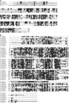
Figure 1.
Figure 1.
Amino Acid Sequence Comparisons of Cr-P4H-1. Alignment of the amino acid residues of Cr-P4H-1A (A) with those of the Arabidopsis P4H-1 (At-P4H-1), the P. bursaria Chlorella virus-1 P4H (PBCV-1 P4H), and the C-terminal region of the human C-P4H α(I) subunit [Hs α(I)] and the Cr-P4H-1 (B) with those of the other putative C. reinhardtii P4Hs. The C-terminal extension present only in the variant Cr-P4H-1B is boxed in (B). The putative C. reinhardtii P4Hs are indicated in (B) by their annotation numbers in the C. reinhardtii genome draft release, version 2.0, of the Department of Energy Joint Genome Institute. The catalytically critical residues are indicated by asterisks. Identical amino acids are shown with black backgrounds.
Figure 2.
Figure 2.
Phylogenetic Tree of Cr-P4H-1 and Other P4Hs. Phylogenetic relationships between the amino acid sequences of Cr-P4H-1A and the C-P4H α subunits from human [Hs α(I), α(II), and α(III)], mouse [Mm α(I), α(II), and α(III)], rat [Rn α(I) and α(III)], chicken [Gg α(I)], C. elegans (Ce PHY-1, PHY-2, and PHY-3), Brugia malayi (Bm PHY-1), Onchocerca volvulus (Ov PHY-1), D. melanogaster [Dm α(I)], and P4Hs from Arabidopsis (At-P4H-1 and At-P4H-2) and PBCV-1 P4H. The neighbor-joining tree was constructed using MEGA, version 2.1 (Kumar et al. 2001). Bootstrap values determined from 1000 replications are shown at each node. Bar = 10% divergence.
Figure 3.
Figure 3.
Expression of Recombinant Cr-P4H-1A and Cr-P4H-1B Polypeptides in Insect Cells or in E. coli and Their Purification. The recombinant Cr-P4H-1A was expressed in insect cells (A) and in E. coli (B), and Cr-P4H-1B was expressed in E. coli (C). Samples of the soluble (lane 1) and insoluble (lane 2) fractions of the cell lysates were analyzed by SDS-PAGE under reducing conditions. The Cr-P4H-1A was purified from insect cells by gel filtration (lane 3 in [A]), and Cr-P4H-1A and Cr-P4H-1B were purified from E. coli using a Ni-NTA Sepharose column (lane 3 in [B] and [C]).
Figure 4.
Figure 4.
CD Analysis of the Temperature Stability of the Recombinant Cr-P4H-1A Polypeptide. Mean residue ellipticity at 222 nm as a function of temperature (T) measured during denaturation from 20 to 90°C. mrw, mean residue weight.
Figure 5.
Figure 5.
Analysis of the Hydroxylation of the Pro Residues in (Pro-Pro-Gly)10 by Cr-P4H-1A. The columns indicate the degree of hydroxylation of the Pro residues in the X- and Y-positions in the X-Y-Gly triplets. P, Pro; G, Gly.
Figure 6.
Figure 6.
Construct for Generating a Double-Stranded RNA Hairpin against the Gene Transcript Encoding Cr-P4H-1 and PCR Analysis of the Presence of the RNAi Construct in Transformed Cells. (A) The sense and antisense strands of part of the coding sequence for Cr-P4H-1 (the antisense long arm is written backward) and the phleomycin resistance gene (ble) are under the control of the C. reinhardtii ribulose bisphosphate carboxylase/oxygenase (RBCS2) promoter. (B) PCR analysis of the presence of the RNAi construct in one control line transformed with the empty vector and one Cr-P4H-1 RNAi line (RNAi-1). Primer pairs 1 and 2 described in Methods were used to amplify 950- and 750-bp regions of the RNAi hairpin construct, respectively, using genomic DNA isolated from the transformed cells as a template. Amplification using ribulose-1,5-bisphosphate carboxylase/oxygenase (Rubisco) primers was used as a control for analysis of the amounts of template DNA.
Figure 7.
Figure 7.
Transmission Electron Micrographs of High-Pressure Frozen and Freeze-Substituted Samples of C. reinhardtii Cells Electroporated with a Double-Stranded RNA Construct against Cr-P4H-1. (A) Wild-type control cells transformed with the empty pSP124S plasmid show a clearly defined, multilayered cell wall. Bar = 1000 nm. (B) The wall is less well defined and uniform in a typical Cr-P4H-1 knockdown cell. Bar = 1000 nm. (C) The distinct cell wall layers (labeled W1, W2, W6, and W7) can be seen in the control cells at higher magnification. Bar = 1000 nm. (D) to (F) The walls of the knockdown cells are seen at higher magnification to lack the multilayered structure, and in some cells, the wall is clearly detached from the plasma membrane. Bar = 100 nm.

References

    1. Abrams, E.W., and Andrew, D.J. (2002). Prolyl 4-hydroxylase α-related proteins in Drosophila melanogaster: Tissue-specific embryonic expression of the 99F8-9 cluster. Mech. Dev. 112 165–171. - PubMed
    1. Adair, W.S., and Snell, W.J. (1990). The Chlamydomonas reinhardtii cell wall: Structure, biochemistry, and molecular biology. In Organization and Assembly of Plant and Animal Extracellular Matrix, W.S. Adair and R.P. Mecham, eds (San Diego, CA: Academic Press), pp. 15–84.
    1. Annunen, P., Autio-Harmainen, H., and Kivirikko, K.I. (1998). The novel type II prolyl 4-hydroxylase is the main enzyme form in chondrocytes and capillary endothelial cells, whereas the type I enzyme predominates in most cells. J. Biol. Chem. 273 5989–5992. - PubMed
    1. Annunen, P., Helaakoski, T., Myllyharju, J., Veijola, J., Pihlajaniemi, T., and Kivirikko, K.I. (1997). Cloning of the human prolyl 4-hydroxylase α subunit isoform α(II) and characterization of the type II enzyme tetramer. The α(I) and α(II) subunits do not form a mixed α(I)α(II)β2 tetramer. J. Biol. Chem. 272 17342–17348. - PubMed
    1. Annunen, P., Koivunen, P., and Kivirikko, K.I. (1999). Cloning of the α subunit of prolyl 4-hydroxylase from Drosophila and expression and characterization of the corresponding enzyme tetramer with some unique properties. J. Biol. Chem. 274 6790–6796. - PubMed

Publication types

MeSH terms

LinkOut - more resources