. 2007 Mar;143(3):1173-88.
doi: 10.1104/pp.106.093435. Epub 2007 Jan 12.

The Arabidopsis aleurone layer responds to nitric oxide, gibberellin, and abscisic acid and is sufficient and necessary for seed dormancy

Affiliations

The Arabidopsis aleurone layer responds to nitric oxide, gibberellin, and abscisic acid and is sufficient and necessary for seed dormancy

Paul C Bethke et al. Plant Physiol. 2007 Mar.

Abstract

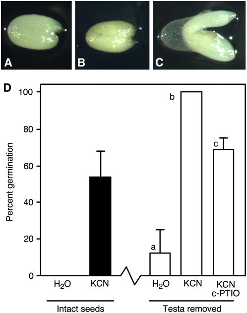
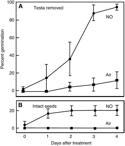
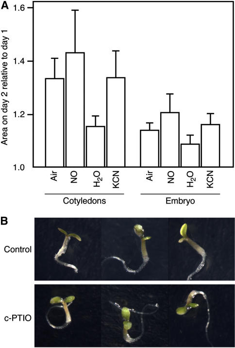
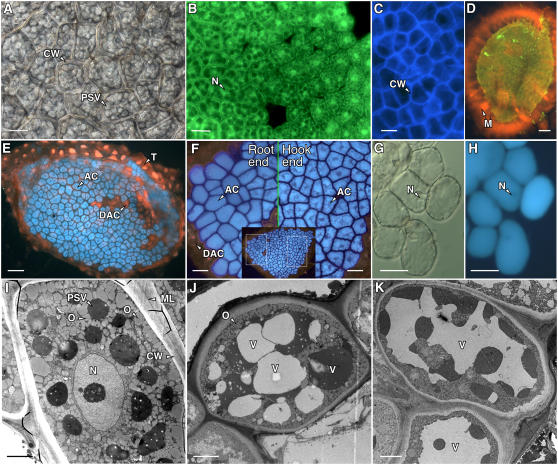
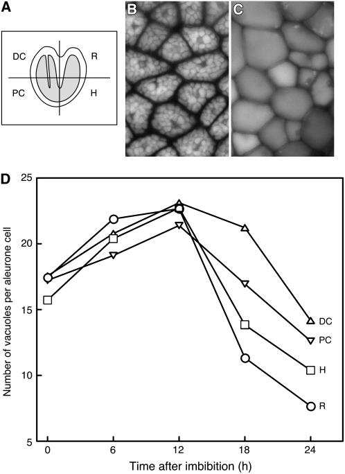
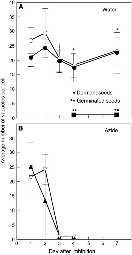
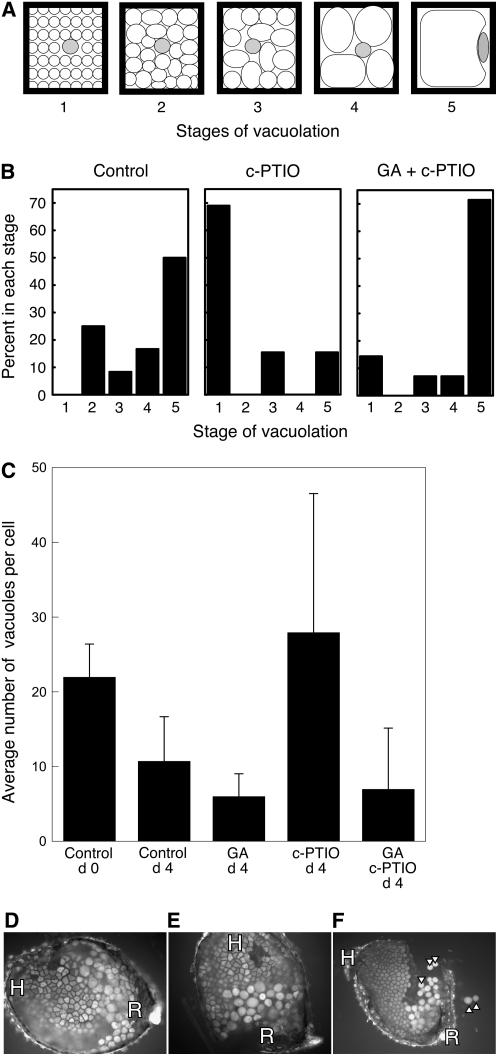
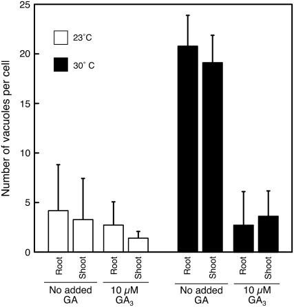
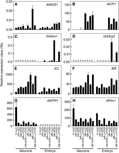
Seed dormancy is a common phase of the plant life cycle, and several parts of the seed can contribute to dormancy. Whole seeds, seeds lacking the testa, embryos, and isolated aleurone layers of Arabidopsis (Arabidopsis thaliana) were used in experiments designed to identify components of the Arabidopsis seed that contribute to seed dormancy and to learn more about how dormancy and germination are regulated in this species. The aleurone layer was found to be the primary determinant of seed dormancy. Embryos from dormant seeds, however, had a lesser growth potential than those from nondormant seeds. Arabidopsis aleurone cells were examined by light and electron microscopy, and cell ultrastructure was similar to that of cereal aleurone cells. Arabidopsis aleurone cells responded to nitric oxide (NO), gibberellin (GA), and abscisic acid, with NO being upstream of GA in a signaling pathway that leads to vacuolation of protein storage vacuoles and abscisic acid inhibiting vacuolation. Molecular changes that occurred in embryos and aleurone layers prior to germination were measured, and these data show that both the aleurone layer and the embryo expressed the NO-associated gene AtNOS1, but only the embryo expressed genes for the GA biosynthetic enzyme GA3 oxidase.


Figures

Figure 1.
Figure 1.
Arabidopsis seeds remain dormant when the testa is removed but germinate when exposed to cyanide vapors. Seeds lacking the testa are shown in A to C 3 d (A) and 28 d (B and C) after testa removal. The seed in C was exposed to KCN vapors for 3 d beginning 25 d after testa removal. D, Germination percentages after 5 d for intact seeds exposed to water vapor or KCN vapor for 2 d, and for seeds with the testa removed exposed to water vapor, KCN vapor, or KCN vapors in the presence of c-PTIO. Each experiment was repeated at least three times with five to 14 seeds per treatment. Bars with different lowercase letters are significantly different with P < 0.05.
Figure 2.
Figure 2.
Germination of Arabidopsis seeds lacking the testa is stimulated by NO gas. Purified NO gas or air were passed over seeds with the testa removed for 1 d (A) or over intact seeds for 2 d (B), and germination was scored every day for 4 d. Data are means ± se for six to 17 seeds.
Figure 3.
Figure 3.
Embryos from dormant C24, Cvi, and Kas2 ecotype Arabidopsis seeds and from nondormant Col ecotype seeds grow when removed from the seed. All embryos were photographed 5 d after isolation. [See online article for color version of this figure.]
Figure 4.
Figure 4.
Embryos from nondormant seeds grow more rapidly than embryos from dormant seeds when imbibed on media containing mannitol or PEG as osmotica. A, Micrographs of embryos 7 d after removal from nondormant or dormant C24 Arabidopsis seeds. B, Surface area of isolated embryos growing on a substrate containing up to 500 mOsmol mannitol or PEG for 2 d relative to the surface area on day 0. C, Relative surface area of cotyledons, hypocotyls, and roots 1 or 2 d after placing freshly isolated embryos from dormant (D) or nondormant (ND) C24 Arabidopsis seeds on 300 mOsmol mannitol or 300 mOsmol PEG.
Figure 5.
Figure 5.
The mean growth potential of embryos from dormant Arabidopsis seeds is not changed significantly following treatment of seeds with NO gas or cyanide vapors, or embryos with the NO scavenger c-PTIO. A, Change in relative surface area of the cotyledons or the whole embryo for embryos removed from seeds exposed to air, NO gas, water vapor, or cyanide vapor for 2 d. Isolated embryos were imbibed on agarose containing 300 mOsmol mannitol. Day 1 is 1 d after the end of the treatment period (n > 10). B, Embryos 5 d after isolation from dormant C24 Arabidopsis seeds. Embryos were placed on water agarose without (control) or with 100 μm c-PTIO. [See online article for color version of this figure.]
Figure 6.
Figure 6.
Cells in the Arabidopsis endosperm are typical aleurone cells. Differential interference contrast (A and G), fluorescence (B–F, H), and electron microscopy (I–K) images of Arabidopsis aleurone layers. A, Region of an aleurone layer showing aleurone cells surrounded by a thick cell wall (CW) and containing numerous PSVs. B, Aleurone layer labeled with the vital fluorescent probe fluorescein diacetate showing green fluorescence from the cytosol and nucleus (N) of live cells. C, Cellulose in the Arabidopsis aleurone cell wall labeled with calcofluor white M2R. D, An extensive mucilage layer (M) that was labeled with orange fluorescent Congo red was retained by the testa cells associated with the isolated aleurone layer. E, Aleurone cells (AC) incubated with monochlorobimane accumulated blue fluorescent bimane in their PSV. The testa and nuclei in dead aleurone cells (DAC) in E fluoresced red after labeling with propidium iodide. F, Cells in Arabidopsis aleurone layers closest to the root became less angular in shape sooner than cells near the hypocotyl hook. Shown are higher magnification images that correspond to the boxed regions of the single aleurone layer shown in the inset. G, High magnification image of individual aleurone cells close to the site of root emergence. Cell walls had begun to separate, and individual cells had become nearly spherical. H, Cells in G are still alive, as indicated by the fluorescence from the vacuole and lack of propidium iodide fluorescence from the nucleus (N). I, Electron micrograph of an aleurone cell 1 h after imbibition showing the numerous PSVs and oleosomes (O). The aleurone cell wall and middle lamella (ML) are also indicated. J and K, Aleurone cells 24 h after imbibition. Note the enlargement of the vacuoles (V) and reduction in the number of oleosomes compared with those in I. The vacuoles in J are heterogeneous with regard to electron density. Scale bars are 2 μm in I to K, 10 μm in A, G, and H, 20 μm in B, C, and F, and 60 μm in D and E.
Figure 7.
Figure 7.
Aleurone cells in nondormant Col Arabidopsis seeds vacuolate prior to germination. A, Schematic of an Arabidopsis seed illustrating the four regions of the aleurone layer where the number of PSVs per cell was quantified. In this illustration, the embryo is shaded in gray. Typical images of aleurone cells shortly after imbibition (B) and shortly after germination (C), where cells in B have many PSVs per cell but cells in C have only one. D, Quantification of PSV number 1, 6, 12, 18, and 24 h after seed imbibition. R, Root; H, hypocotyl; PC, proximal end of cotyledons; DC, distal end of cotyledons. Data in D are means of 32 to 266 cells. sds in D range from 4.21 to 7.19, and error bars have been omitted for clarity.
Figure 8.
Figure 8.
Aleurone cells vacuolate in seeds that germinate. Mildly dormant C24 Arabidopsis seeds were imbibed in water (A) or exposed to 10 μm NaN3 (B), and the number of PSV per aleurone cell was determined. Most of the seeds in A were dormant, but some were not and germinated by 96 h. All of the seeds in B germinated by 96 h. White and black symbols represent the aleurone layer adjacent to the root/hypocotyl and cotyledons, respectively. Note that vacuolation of aleurone cells was tightly correlated with dormancy loss.
Figure 9.
Figure 9.
Vacuole number per cell is a dynamic parameter in aleurone layers incubated in vitro. The average number of vacuoles per cell between 0 and 4 d after removal of the aleurone layer from an imbibed seed is shown in A for the Col, Kas2, and Cvi ecotypes, and in B for the C24 ecotype of Arabidopsis. The Col ecotype seeds were nondormant, and the Kas2, Cvi, and C24 ecotype seeds were dormant. Aleurone layers in B were incubated on agarose with and without 10 nm ABA. In both A and B, data are presented for that part of the aleurone layer proximal to the root/hypocotyl (root side) and that part proximal to the cotyledons (shoot side).
Figure 10.
Figure 10.
The NO scavenger c-PTIO inhibits vacuolation of aleurone cells in isolated, wild-type Arabidopsis aleurone layers but not in layers from the Spy-1 mutant. A, The extent of vacuolation for each layer in B was scored based on the stage (1–5) of vacuolation typical for the cells in the layer. A single cell is diagrammed for each stage. B, Vacuolation was delayed in C24 aleurone layers treated with 50 μm c-PTIO, and the effect of c-PTIO was reversed by 10 μm GA3. The data are from a single, representative experiment. C, Vacuolation of cells in aleurone layers from nondormant Ler seeds was stimulated by 10 μm GA3 and inhibited by c-PTIO. Data are means ± sd. D to F, Micrographs of isolated aleurone layers from the root/hypocotyl side of Spy-1 mutant seed showing that cells originally near the root (R) but not the hypocotyl (H) exhibit evidence of vacuolation and wall weakening in the presence of no additions (D) or 100 μm c-PTIO (E and F). Note that the walls of some cells in F were weakened enough that individual cells were released from the layer (arrowheads).
Figure 11.
Figure 11.
GA promotes the vacuolation of cells in isolated Col aleurone layers incubated at 30°C. Layers were incubated for 2 d in vitro on agarose with or without 10 μm GA3 and at 23°C or 30°C. Note that cells did not vacuolate at 30°C in the absence of GA but did vacuolate at 30°C in the presence of GA. Data are presented for that part of the aleurone layer proximal to the root/hypocotyl (root) and that part proximal to the cotyledons (shoot). Bars are means ± sd.
Figure 12.
Figure 12.
Quantification of relative mRNA abundance for genes associated with dormancy or germination in the aleurone layer and embryo of Arabidopsis seeds. Dormant seeds of C24 Arabidopsis were imbibed on water agarose supplemented with or without 200 μm c-PTIO and exposed to water vapors or KCN (300 μm) vapors. RNA abundance in A to H was quantified using qPCR for samples isolated 1, 24, or 48 h after imbibition and is expressed relative to the abundance of reference genes. Asterisks indicate samples where the gene product was not detected.

Similar articles

Cited by

References

    1. Baskin J, Baskin C (1983) Seasonal changes in germination responses of buried seeds of Arabidopsis thaliana and ecological interpretations. Bot Gaz 144 540–543
    1. Bethke PC, Badger MR, Jones RL (2004. a) Apoplastic synthesis of nitric oxide by plant tissues. Plant Cell 16 332–341 - PMC - PubMed
    1. Bethke PC, Gubler F, Jacobsen JV, Jones RL (2004. b) Dormancy of Arabidopsis seeds and barley grains can be broken by nitric oxide. Planta 219 847–855 - PubMed
    1. Bethke PC, Libourel IGL, Jones RL (2006. a) Nitric oxide reduces seed dormancy in Arabidopsis. J Exp Bot 57 517–526 - PubMed
    1. Bethke PC, Libourel IGL, Reinohl V, Jones RL (2006. b) Sodium nitroprusside, cyanide, nitrite, and nitrate break Arabidopsis seed dormancy in a nitric oxide-dependent manner. Planta 223 805–812 - PubMed

Publication types

MeSH terms

LinkOut - more resources