. 2007 Mar;6(3):454-64.
doi: 10.1128/EC.00283-06. Epub 2007 Jan 12.

De novo sphingolipid synthesis is essential for viability, but not for transport of glycosylphosphatidylinositol-anchored proteins, in African trypanosomes

Affiliations

De novo sphingolipid synthesis is essential for viability, but not for transport of glycosylphosphatidylinositol-anchored proteins, in African trypanosomes

Shaheen S Sutterwala et al. Eukaryot Cell. 2007 Mar.

Abstract

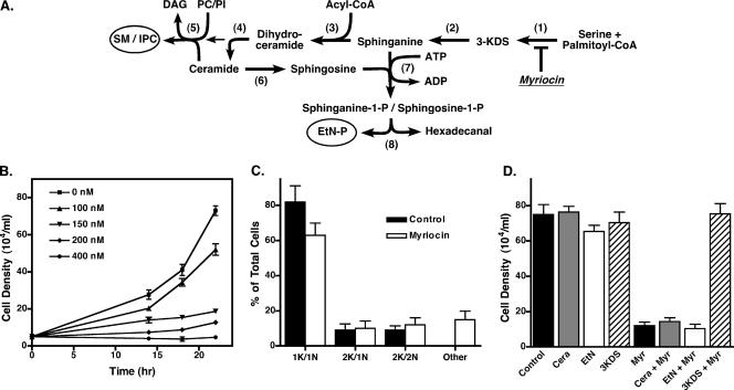
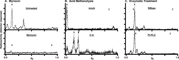
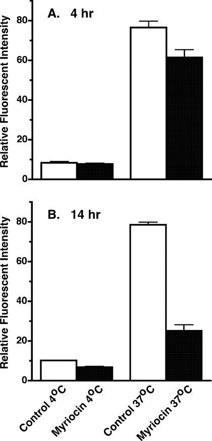
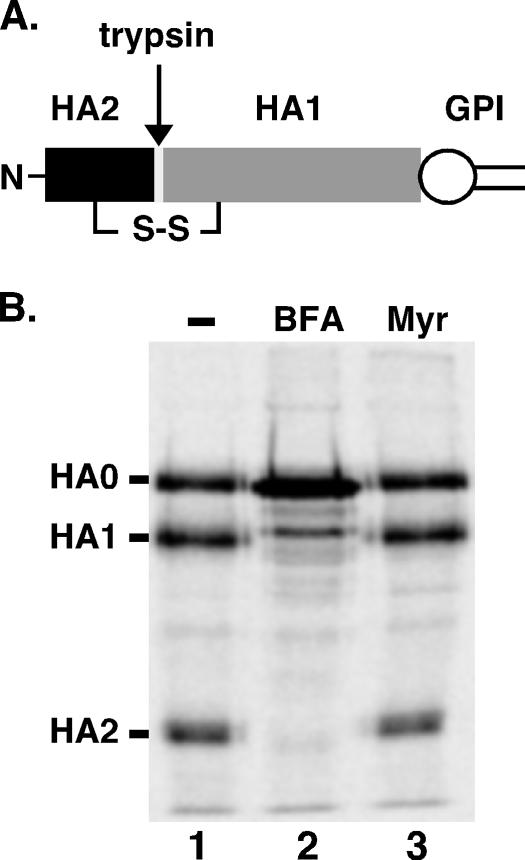
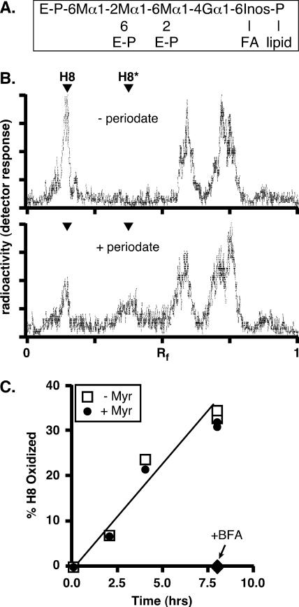
De novo sphingolipid synthesis is required for the exit of glycosylphosphatidylinositol (GPI)-anchored membrane proteins from the endoplasmic reticulum in yeast. Using a pharmacological approach, we test the generality of this phenomenon by analyzing the transport of GPI-anchored cargo in widely divergent eukaryotic systems represented by African trypanosomes and HeLa cells. Myriocin, which blocks the first step of sphingolipid synthesis (serine + palmitate --> 3-ketodihydrosphingosine), inhibited the growth of cultured bloodstream parasites, and growth was rescued with exogenous 3-ketodihydrosphingosine. Myriocin also blocked metabolic incorporation of [3H]serine into base-resistant sphingolipids. Biochemical analyses indicate that the radiolabeled lipids are not sphingomyelin or inositol phosphorylceramide, suggesting that bloodstream trypanosomes synthesize novel sphingolipids. Inhibition of de novo sphingolipid synthesis with myriocin had no adverse effect on either general secretory trafficking or GPI-dependent trafficking in trypanosomes, and similar results were obtained with HeLa cells. A mild effect on endocytosis was seen for bloodstream trypanosomes after prolonged incubation with myriocin. These results indicate that de novo synthesis of sphingolipids is not a general requirement for secretory trafficking in eukaryotic cells. However, in contrast to the closely related kinetoplastid Leishmania major, de novo sphingolipid synthesis is essential for the viability of bloodstream-stage African trypanosomes.


Figures

FIG. 1.
FIG. 1.
Cell growth is inhibited by myriocin. (A) The sphingoid lipid pathway. Enzymes are indicated by numbers in parentheses as follows: (1), serine palmitoyltransferase; (2), 3-ketosphingosine reductase; (3), dihydroceramide synthase;(4), dihydroceramide desaturase; (5), “sphingolipid” synthase; (6), ceramidase; (7), sphingosine kinase; (8), sphingosine-1-phosphate lyase. Final end products are sphingomyelin (SM), inositol phosphorylceramide (IPC), and ethanolamine phosphate (EtN-P). Other abbreviations are 3-ketodihydrosphingosine (3-KDS), phosphatidylcholine (PC), phosphatidylinositol (PI), and diacylglycerol (DAG). The site of inhibition by myriocin is indicated. (B) Bloodstream cell cultures were seeded at 1 × 10⁵ cells/ml in the presence of the indicated concentrations of myriocin, and densities were counted at the indicated times. Means ± standard deviations of triplicate cultures were determined. (C) Fixed and permeabilized bloodstream cells were stained with DAPI to reveal the localization of the nucleus and kinetoplast. The numbers of nuclei and kinetoplasts per cell were counted (n = 100) for myriocin-treated (200 nM, 18 h) and control cultures. Data are means ± standard deviations from three independent experiments. (D) Bloodstream trypanosomes were seeded at 1 × 10⁵ cells/ml and grown for 18 h in the presence or absence of 200 nM myriocin with additional supplementation with either 2.5 μM 3-KDS, 10 μM ceramide (Cera), or 100 μM ethanolamine (EtN). Values are means ± standard deviations from triplicate cultures.
FIG. 2.
FIG. 2.
Myriocin inhibition of de novo sphingolipid synthesis in trypanosomes. (A) Bloodstream-stage trypanosomes grown for 4 h in the presence (bottom) or absence (top) of 200 nM myriocin were pulse-radiolabeled with 50 μCi/ml of [3H]serine for 4 h. Base-resistant lipids were extracted as described in Materials and Methods and analyzed by TLC. The mobilities of sphingomyelin (Rf, 0.12) and ceramide (Rf, 0.89) standards were determined by iodine staining (arrowheads, bottom panel only). (B) Saponified [3H]serine-labeled lipids were subjected to mock treatment (top) or acid methanolysis (bottom) (2 h) as described in Materials and Methods and then analyzed by TLC. Ceramide was added as an internal marker/substrate prior to hydrolysis. The mobilities of unhydrolyzed ceramide (open arrowhead) (Rf, 0.83), the sphingosine product (shaded arrowhead) (Rf, 0.25), and the sphinganine product (solid arrowhead) (Rf, 0.15) are indicated. (C) Base-resistant [3H]serine-labeled lipids from untreated bloodstream trypanosomes were either mock treated (not shown) or digested with either sphingomyelinase (SMase) (top) or bacterial PI-PLC (bottom) with internal sphingomyelin and PI controls, respectively. Samples were prepared and analyzed as for panel A. Solid arrowheads indicate relative mobilities of internal sphingomyelin (top) (Rf, 0.14) and PI (bottom) (Rf, 0.22) control substrates (from the mock treatments), and open arrowheads indicate the respective ceramide (top) (Rf, 0.9) and diacylglycerol (bottom) (Rf, 0.8) products. Note that the solvent systems used for panels A and C are different from that for panel B.
FIG. 3.
FIG. 3.
Myriocin does not block secretory transport in trypanosomes. (A) Bloodstream-form trypanosomes constitutively expressing the soluble secretory reporter BiPN were cultured for 14 h in the absence (−) or presence (+) of 200 nM myriocin (myr.). Cells were pulse-radiolabeled (15 min) with [35S]Met-Cys and then chased for the indicated times in the continued presence or absence of the inhibitor. The BiPN reporter was immunoprecipitated from cell and medium fractions and analyzed by SDS-PAGE and phosphorimaging. The mobilities of endogenous BiP and VSG, and of the BiPN reporter, are indicated on the left. All lanes contain 5 ×10⁶ cell equivalents. Note that the time points used in this assay fall in the linear range for BiPN transport in bloodstream cells (60). (B) Bloodstream cells were cultured for 14 h in the presence or absence of 200 nM myriocin. Cells were then pulse-radiolabeled (3 min) with [35S]Met-Cys and chased in the continued presence (myr) or absence (control [cont]) of the inhibitor. At the indicated times, samples were washed and subjected to hypotonic lysis as described in Materials and Methods, and VSG polypeptides were immunoprecipitated from the released fractions. VSG was also immunoprecipitated from total-cell fractions at the beginning and end of the chase period (T0 and T30), and all samples were analyzed as for panel A. The recovery of released VSG as a fraction of total VSG (average of T0 and T30) is given for each lane. All lanes contain 2 × 10⁶ cell equivalents.
FIG. 4.
FIG. 4.
Myriocin disrupts lysosomal morphology in trypanosomes. Bloodstream-form trypanosomes were cultured for 14 h in the presence or absence of myriocin (200 nM). Fixed/permeabilized control (A to D) and myriocin-treated (14 h) (E to H) cells were stained with anti-BiP as an ER marker (green) (B and F) or anti-p67 as a lysosomal marker (red) (C and G) and were visualized by epifluorescence microscopy. Alternatively, control (I to L) and myriocin-treated (M to P) cells were allowed to endocytose FITC-TL (J and N) for 1 h and were then processed for staining with anti-p67 (K and O). (A, E, I, and M) Merged DIC and DAPI images; (D, H, L, and P) merged three-channel fluorescent images. DAPI staining (blue) reveals nucleus (n) and kinetoplast (k) localization (indicated in panel A only).
FIG. 5.
FIG. 5.
Ultrastructure of myriocin-treated trypanosomes. Cultured bloodstream cells were first pulse-loaded (2 h, 37°C) with colloidal gold (5 nm) coupled to bovine holotransferrin to label the terminal lysosome. Cells were then cultured in the absence (A and B) or presence (C and D) of myriocin (200 nM) for 14 h and processed as described for electron microscopy. (A to D) Representative cell sections. Bar, 1.0 μm. White arrowheads indicate lysosomes containing colloidal gold particles. cont, control; myr, myriocin. (E to H) Corresponding images in the lysosomal region taken at a higher magnification. Bar, 0.2 μm.
FIG. 6.
FIG. 6.
Myriocin affects endocytic transport in trypanosomes. Bloodstream cells were cultured for 4 h (A) or 14 h (B) in the presence or absence of 200 nM myriocin. Cells were then incubated at 4°C or 37°C for 1 h in a medium containing 20 μg/ml FITC-tomato lectin, washed, and analyzed by flow cytometry. Uptake is presented as relative fluorescent intensity, in arbitrary units. Values are means ± standard deviations from three independent experiments.
FIG. 7.
FIG. 7.
Transport of HA-GPI to the plasma membranes of HeLa cells is unaffected by myriocin treatment. (A) Schematic representation of HA-GPI. The protein is synthesized as a GPI-anchored polypeptide (HA0, corresponding to the ectodomain of influenza virus hemagglutinin protein) containing a disulfide bond as shown. Upon trypsin treatment, HA0 yields two disulfide-linked fragments, HA2 and HA1. (B) HeLa cells expressing HA-GPI were preincubated for 30 min with myriocin (125 μM) or BFA (5 μg/ml) as indicated and were then pulse-labeled with 200 μCi/ml [35S]methionine-cysteine for 20 min. The cells were chased for 2 h in the presence of drugs before being trypsinized to convert cell surface HA-GPI molecules into HA2 and HA1. The extent of conversion of radiolabeled protein to HA2 and HA1 was determined after immunoprecipitation and SDS-PAGE. A scan of a typical fluorography is presented. No HA2 is seen in the BFA-treated cells, whereas radiolabeled HA2 is readily observed in both control (−) and myriocin-treated cells.
FIG. 8.
FIG. 8.
Transport of free GPIs to the cell surface in HeLa cells is unaffected by myriocin. (A) Structure of the mammalian GPI H8, showing glycosidic linkages and sites of ethanolamine-phosphate (E-P) attachment. M, mannose; G, glucosamine; Inos, inositol; FA, fatty acid. (B) HeLa cells were first preincubated in a low-glucose medium with 6 μg/ml tunicamycin for 30 min at 37°C, then metabolically radiolabeled with [2-3H]mannose for 2.5 h at 37°C, and finally chased for 18 h in complete medium. At the end of the chase period, the cells were washed, incubated for 30 min on ice in PBS (without [−] or with [+] 10 mM sodium metaperiodate), and washed again in PBS supplemented with 150 mM glycerol. The cells were collected, and lipids were extracted and analyzed by TLC (chloroform-methanol-water, 10:10:3 [vol/vol/vol]). [3H]mannose-labeled H8 migrates with an Rf of ∼0.25 (the identity of H8 was confirmed by establishing its sensitivity to hydrolysis by GPI-specific phospholipase D [not shown]). Periodate treatment (lower panel) converts ∼50% of H8 to oxidized H8 (H8*). Several faster-migrating radiolabeled peaks were unaffected by periodiate, suggesting that these unidentified species are intracellular. (C) HeLa cells were first preincubated with myriocin (25 μg/ml) for 20 min, then pulse-labeled with [2-3H]mannose, and finally chased for 8 h in complete medium containing 62.5 μM myriocin (+ Myr). Control (untreated) (− Myr) and BFA-treated samples (cells were preincubated with BFA [2.5 μg/ml] and chased in the presence of the drug) were analyzed in parallel. Cells were taken at different time points and subjected to periodate oxidation to determine the fraction of radiolabeled H8 that was transported from the ER and located at the cell surface.

Similar articles

Cited by

References

    1. Agusti, R., A. S. Couto, O. Campetella, A. C. C. Frasch, and R. M. de Lederkremer. 1998. Structure of the glycosylphosphatidylinositol-anchor of the trans-sialidase from Trypanosoma cruzi metacyclic trypomastigote forms. Mol. Biochem. Parasitol. 97:123-131. - PubMed
    1. Alexander, D. L., K. J. Schwartz, A. E. Balber, and J. D. Bangs. 2002. Developmentally regulated trafficking of the lysosomal membrane protein p67 in Trypanosoma brucei. J. Cell Sci. 115:3253-3263. - PubMed
    1. Bagnat, M., S. Keränen, A. Shevchenko, A. Shevchenko, and K. Simons. 2000. Lipid rafts function in biosynthetic delivery of proteins to the cell surface in yeast. Proc. Natl. Acad. Sci. USA 97:3254-3259. - PMC - PubMed
    1. Bangs, J. D., N. Andrews, G. W. Hart, and P. T. Englund. 1986. Posttranslational modification and intracellular transport of a trypanosome variant surface glycoprotein. J. Cell Biol. 103:255-263. - PMC - PubMed
    1. Bangs, J. D., E. M. Brouch, D. M. Ransom, and J. L. Roggy. 1996. A soluble secretory reporter system in Trypanosoma brucei: studies on endoplasmic reticulum targeting. J. Biol. Chem. 271:18387-18393. - PubMed

Publication types

MeSH terms

LinkOut - more resources