. 2007 Feb;39(2):168-77.
doi: 10.1038/ng1943. Epub 2007 Jan 14.

The neuronal sortilin-related receptor SORL1 is genetically associated with Alzheimer disease

Affiliations

The neuronal sortilin-related receptor SORL1 is genetically associated with Alzheimer disease

Ekaterina Rogaeva et al. Nat Genet. 2007 Feb.

Abstract

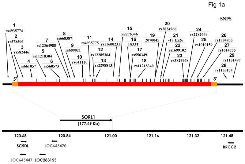
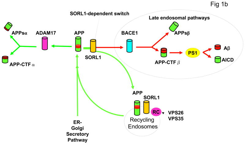
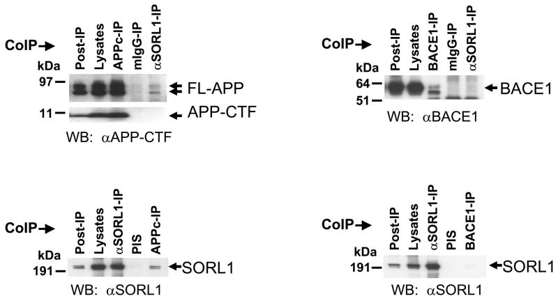
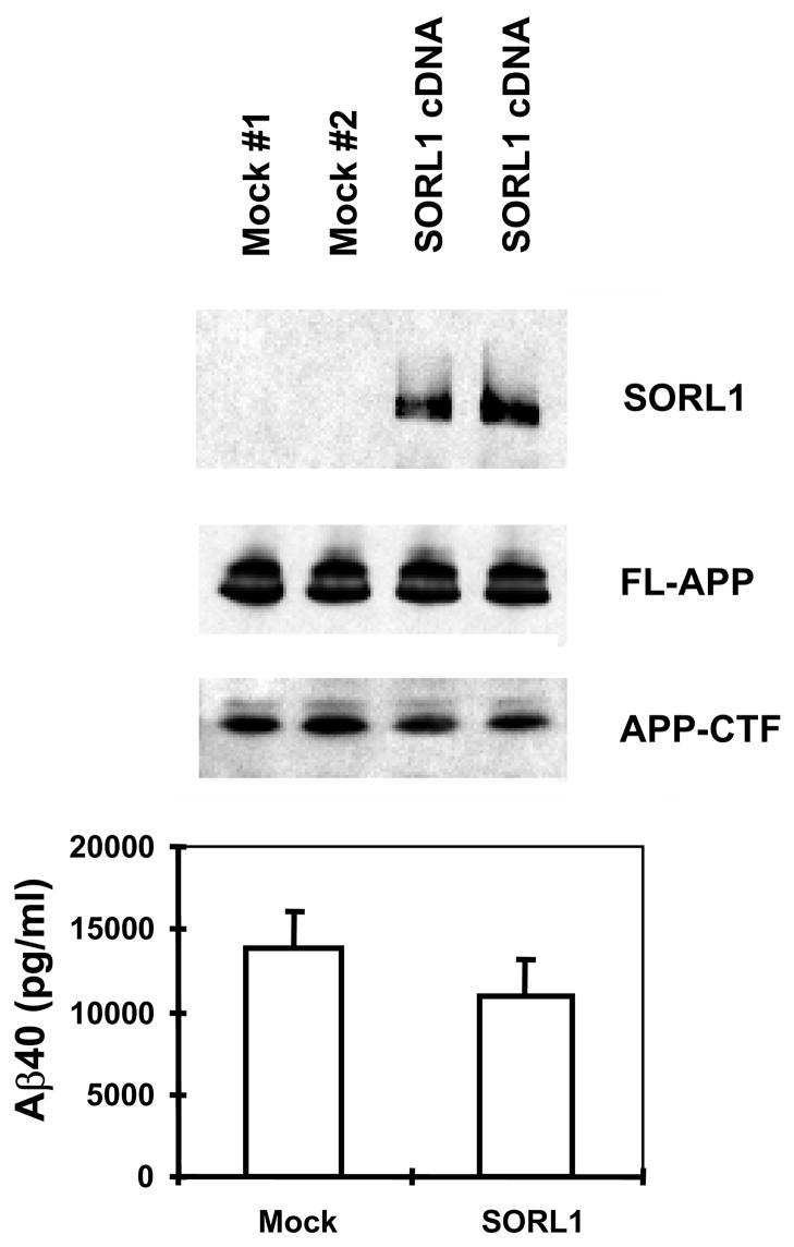
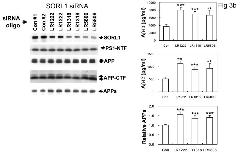
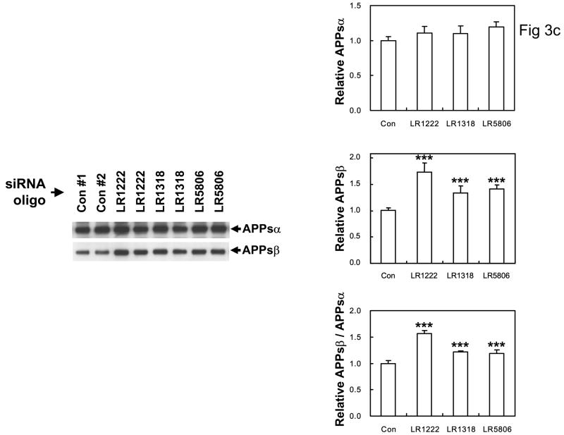
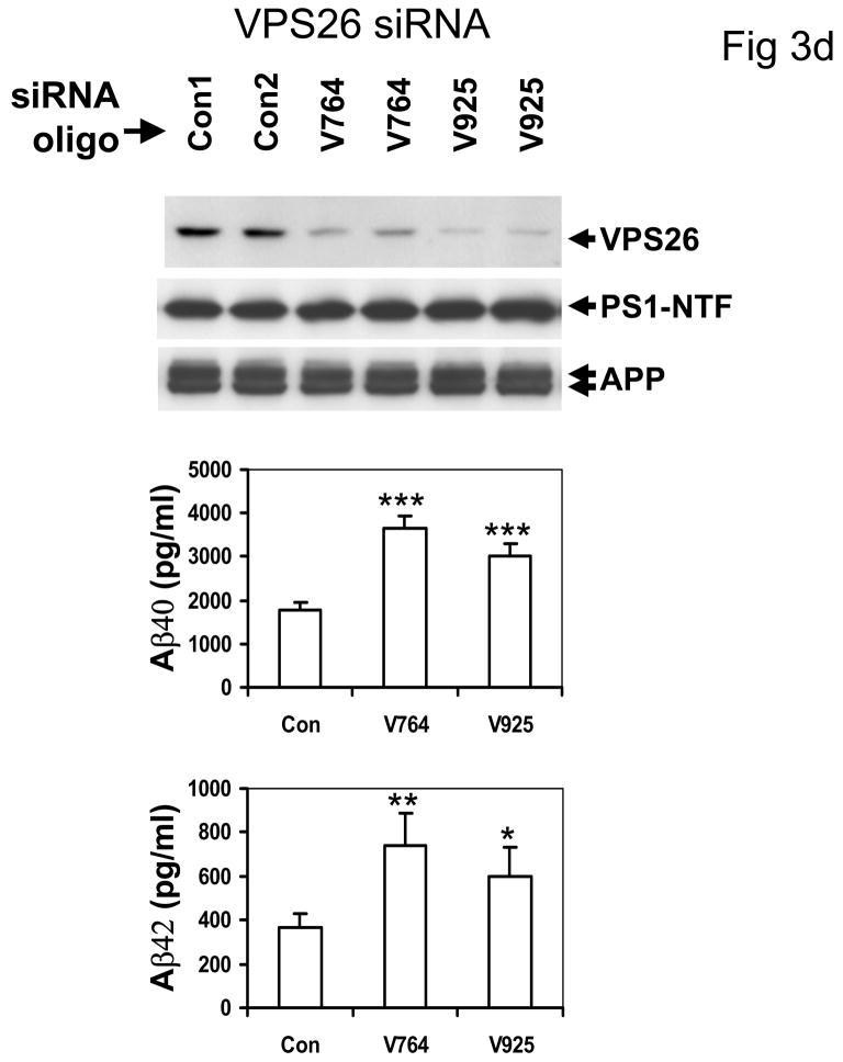
The recycling of the amyloid precursor protein (APP) from the cell surface via the endocytic pathways plays a key role in the generation of amyloid beta peptide (Abeta) in Alzheimer disease. We report here that inherited variants in the SORL1 neuronal sorting receptor are associated with late-onset Alzheimer disease. These variants, which occur in at least two different clusters of intronic sequences within the SORL1 gene (also known as LR11 or SORLA) may regulate tissue-specific expression of SORL1. We also show that SORL1 directs trafficking of APP into recycling pathways and that when SORL1 is underexpressed, APP is sorted into Abeta-generating compartments. These data suggest that inherited or acquired changes in SORL1 expression or function are mechanistically involved in causing Alzheimer disease.


Conflict of interest statement

COMPETING INTERESTS

The authors have no competing financial interests.

Figures

Figure 1
Figure 1
Genomic map of SORL1 gene showing the location of SNPs genotyped in this study. Orange bars represent the 5′UTR and 3′UTR, red bar represents intragenic regions, vertical bars represent each of the 48 exons. SNPs 1, 28 and 29 are located in extragenic intervals. B: Diagram of APP processing pathways. APP holoprotein is synthesized in the endoplasmic reticulum (ER) and Golgi. Proteolytic cleavage through the Aβ peptide domain by ADAM17 and other α-secretase enzymes generates N-terminal soluble APPsα and membrane-bound APP-CTFα fragments. Sequential cleavage by BACE1 (β-secretase) generates N-terminal APPsβ and membrane bound APP-CTFβ fragments. The latter undergoes presenilin-dependent γ-secretase cleavage to generate Aβ and amyloid intracellular domain (AICD). SORL1 binds both APP holoprotein (see Fig. 3) and VPS35 (not shown) and acts as a sorting receptor for APP holoprotein. Absence of SORL1 switches APP holoprotein away from the retromer recycling pathway, and instead directs APP into the β-secretase cleavage pathway, increasing APPsβ production (Fig 2c) and then into the γ-secretase cleavage pathway to generate Aβ (see Fig. 2b). Blockade of the retromer complex (RC) by inhibiting retromer complex proteins such as VPS26 (Fig. 2d) or VPS35 has a similar effect, also increasing APPsβ and Aβ production.
Figure 1
Figure 1
Genomic map of SORL1 gene showing the location of SNPs genotyped in this study. Orange bars represent the 5′UTR and 3′UTR, red bar represents intragenic regions, vertical bars represent each of the 48 exons. SNPs 1, 28 and 29 are located in extragenic intervals. B: Diagram of APP processing pathways. APP holoprotein is synthesized in the endoplasmic reticulum (ER) and Golgi. Proteolytic cleavage through the Aβ peptide domain by ADAM17 and other α-secretase enzymes generates N-terminal soluble APPsα and membrane-bound APP-CTFα fragments. Sequential cleavage by BACE1 (β-secretase) generates N-terminal APPsβ and membrane bound APP-CTFβ fragments. The latter undergoes presenilin-dependent γ-secretase cleavage to generate Aβ and amyloid intracellular domain (AICD). SORL1 binds both APP holoprotein (see Fig. 3) and VPS35 (not shown) and acts as a sorting receptor for APP holoprotein. Absence of SORL1 switches APP holoprotein away from the retromer recycling pathway, and instead directs APP into the β-secretase cleavage pathway, increasing APPsβ production (Fig 2c) and then into the γ-secretase cleavage pathway to generate Aβ (see Fig. 2b). Blockade of the retromer complex (RC) by inhibiting retromer complex proteins such as VPS26 (Fig. 2d) or VPS35 has a similar effect, also increasing APPsβ and Aβ production.
Figure 2
Figure 2
A: Small quantities of endogenous APP holoprotein but not APP C-terminal fragments (APP-CTFs, generated by α- or β-secretase) can be co-immunoprecipitated with endogenous SORL1 (Top panel). Conversely small quantities of endogenous SORL1 can be co-precipitated with endogenous APP holoprotein (Bottom panel). B: SORL1 does not interact with BACE1 (β-secretase). Co-immunoprecipitations with antibodies to over-expressed BACE1-V5 fail to capture SORL1 (Bottom panel). Conversely, SORL1-directed antibodies do not co-immunoprecipitate BACE1 (Top panel) even though BACE1 also traffics through the endosome to Golgi pathway.
Figure 3
Figure 3
A: Over-expression of SORL1 reduces Aβ40 (and Aβ42 not shown) secretion (p < 0.05). Upper panel: Representative data of Western blot for SORL1 and APP in HEK293 cells stably expressing APPSwe, and transiently transfected with empty vector (mock) or SORL1 (n = 2 independent transfections). Lower panel: Bar charts of ELISA assays of secreted Aβ40 (and Aβ42 not shown) following SORL1 over-expression. Error bar: SD; *p<0.05 compared to Control (2-tailed t-test); n = 2 replications. B: Left panel: Suppression of SORL1 expression with three independent siRNA primers (LR1222, LR1318, and LR5806) did not alter the expression levels or maturation of APP, APP-C83 C-terminal fragments or PS1, but (Right panel) significantly increased Aβ40 and Aβ42 secretion and APPs secretion (*p <0.005, ** p < 0.001 2-tailed t-test compared to controls, n = 5 replications, 3 siRNAi oligomers). C: anti-SORL1 siRNA treatment results in significant increases in APPsβ secreted into the media, but no significant change in APPsα levels. Left panel: Western blots of conditioned media from cells treated with nonsense siRNA oligo-nucleotides (Controls #1 and #2) or with anti-SORL1 siRNA oligonucleotides investigated with the 2H3 antibody to APPsα or with SW192 antibody to APPsβ (n = 5 replications). Right panel: quantitation normalized to the control. ** p < 0.0001 2-tailed t-test compared to controls, n = 5 replications. D: Top panel: suppression of VPS26, another member of the VPS10 family involved in the retromer pathways also did not alter APP or PS1 maturation, but (Middle and Bottom panels) did increase both Aβ40 and Aβ42 secretion (*p <0.005, ** p < 0.001 2-tailed t-test compared to controls, n = 5 replications, 2 siRNA oligomers). The control primer had no such effect.
Figure 3
Figure 3
A: Over-expression of SORL1 reduces Aβ40 (and Aβ42 not shown) secretion (p < 0.05). Upper panel: Representative data of Western blot for SORL1 and APP in HEK293 cells stably expressing APPSwe, and transiently transfected with empty vector (mock) or SORL1 (n = 2 independent transfections). Lower panel: Bar charts of ELISA assays of secreted Aβ40 (and Aβ42 not shown) following SORL1 over-expression. Error bar: SD; *p<0.05 compared to Control (2-tailed t-test); n = 2 replications. B: Left panel: Suppression of SORL1 expression with three independent siRNA primers (LR1222, LR1318, and LR5806) did not alter the expression levels or maturation of APP, APP-C83 C-terminal fragments or PS1, but (Right panel) significantly increased Aβ40 and Aβ42 secretion and APPs secretion (*p <0.005, ** p < 0.001 2-tailed t-test compared to controls, n = 5 replications, 3 siRNAi oligomers). C: anti-SORL1 siRNA treatment results in significant increases in APPsβ secreted into the media, but no significant change in APPsα levels. Left panel: Western blots of conditioned media from cells treated with nonsense siRNA oligo-nucleotides (Controls #1 and #2) or with anti-SORL1 siRNA oligonucleotides investigated with the 2H3 antibody to APPsα or with SW192 antibody to APPsβ (n = 5 replications). Right panel: quantitation normalized to the control. ** p < 0.0001 2-tailed t-test compared to controls, n = 5 replications. D: Top panel: suppression of VPS26, another member of the VPS10 family involved in the retromer pathways also did not alter APP or PS1 maturation, but (Middle and Bottom panels) did increase both Aβ40 and Aβ42 secretion (*p <0.005, ** p < 0.001 2-tailed t-test compared to controls, n = 5 replications, 2 siRNA oligomers). The control primer had no such effect.

Similar articles

Cited by

References

    1. Mattson MP. Pathways towards and away from Alzheimer’s disease. Nature. 2004;430:631–9. - PMC - PubMed
    1. Goate AM, et al. Segregation of a missense mutation in the amyloid precursor protein gene with Familial Alzheimer Disease. Nature. 1991;349:704–706. - PubMed
    1. Sherrington R, et al. Cloning of a gene bearing missense mutations in early onset familial Alzheimer’s disease. Nature. 1995;375:754–760. - PubMed
    1. Rogaev EI, et al. Familial Alzheimer’s disease in kindreds with missense mutations in a novel gene on chromosome 1 related to the Alzheimer’s Disease type 3 gene. Nature. 1995;376:775–778. - PubMed
    1. Saunders A, et al. Association of Apoliprotein E allele e4 with the late-onset familial and sporadic Alzheimer Disease. Neurology. 1993;43:1467–1472. - PubMed

Publication types