. 2007 Feb;150(4):424-33.
doi: 10.1038/sj.bjp.0707123. Epub 2007 Jan 15.

Long-term treatment with TGFbeta1 impairs mechanotransduction in bovine aortic endothelial cells

Affiliations

Long-term treatment with TGFbeta1 impairs mechanotransduction in bovine aortic endothelial cells

M Watanabe et al. Br J Pharmacol. 2007 Feb.

Abstract

Background and purpose: Vascular endothelial cells play a role in the physiological response to mechanical stress. Transforming growth factor beta1 (TGFbeta1) induces morphological changes in endothelial cells, and this may alter their mechanosensitive responses. The aim of this study was to examine the effects of TGFbeta1 on hypotonic stress (HTS)-induced responses in bovine aortic endothelial cells (BAECs).

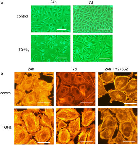
Experimental approach: Cultured BAECs were treated with 3 ng ml(-1) TGFbeta1 for 24 h (24h-TGFbeta1) or 7 days (7d-TGFbeta1). Cytosolic actin fibres were stained with rhodamine-phalloidin. Intracellular Ca2+ concentration was measured using fura2. Tyrosine phosphorylation and RhoA expression were assessed by Western blotting. Expression of RhoA mRNA was assessed by real-time PCR.

Key results: BAECs developed pseudopod-like processes within 24 h and showed a fibroblast-like appearance after 7 days. HTS induced Ca2+ transients via endogenous ATP release in both control and 24h-TGFbeta1 BAECs but not in 7d-TGFbeta1 BAECs. We have previously shown that HTS-induced ATP release is mediated by sequential activation of RhoA and tyrosine kinases. The basal amount of membrane-bound RhoA was significantly lower in 7d-TGFbeta1 than in 24h-TGFbeta1 or control BAECs. HTS increased the membrane-bound RhoA to the same fractional level in 24h-TGFbeta1 and control BAECs, but its net maximal amount was significantly lower in 7d-TGFbeta1. HTS-induced downstream signals of RhoA activation, i.e. the tyrosine phosphorylation of FAK and paxillin, were markedly suppressed in 7d-TGFbeta1 BAECs.

Conclusions and implications: These results indicate that long-term treatment with TGFbeta1 does not impair mechanoreception in BAECs but impairs mechanotransduction by affecting RhoA membrane translocation.


Figures

Figure 1
Figure 1
TGFβ1-induced morphological changes in BAECs. (a) Cells were seeded on culture plates, and cultured for 24 h or 7 days in the absence (control) or presence of 3 ng ml−1 TGFβ1. Transillumination microscopic pictures are shown. Scale bars, 50 μm. (b) Actin fibres were visualized with rhodamine-phalloidin in control and 3 ng ml−1 TGFβ1-treated BAECs after culturing for 24 h or 7days. Effects of 10 μM Y27632, a Rho-kinase inhibitor, are also depicted. Scale bars, 20 μm.
Figure 2
Figure 2
Ca2+ transients in control and TGFβ1-treated BAECs. (a) BAECs were seeded on coverslips and cultured for 7 days. Control cells were left untreated (i). Cells to which 3ng ml−1 TGFβ1 was applied at day 6 and cultured for subsequent 24 h are denoted as 24 h-TGFβ1 (ii). Cells that were cultured in the presence of 3 ng ml−1 TGFβ1 for 7 days are denoted as 7d-TGFβ1 (iii, iv). The effects of HTS (−30%; iiii) and ATP (0.1 μM; iv) on [Ca2+]i were then examined. Representative Ca2+ traces are shown. (b) Hypotonicity-dependence of the HTS-induced Ca2+ transients. The percentage of cells showing HTS-induced Ca2+ transients in a microscopic field was calculated. Each point was obtained from six measurements; vertical lines shown s.e.m. **P<0.01, *P<0.05 vs control.
Figure 3
Figure 3
Effects of TGFβ1 on the expression of RhoA in BAECs in isotonic conditions. (a) BAECs were seeded on culture plates and cultured for 7 days under various conditions. Untreated cells were cultured for 7days with a medium change at day 4. Other cells were cultured for 7 days with an additional medium exchange without (denoted as cont.) or with 3 ng ml−1 TGFβ1 6 h after seeding (denoted as 7d) or 1 or 24 h before cell lysis (denoted as 1 and 24 h, respectively). Cell lysate was then centrifuged at 100 000 g for 1 h, and pellet and supernatant were collected as membrane (upper panel) and cytosolic (lower panel) fractions, respectively. RhoA/β-actin ratio was determined densitometrically and expressed relative to control values in untreated cells. Note that RhoA in the membrane fraction but not in the cytosolic fraction was decreased only in 7d-TGFβ1. **P<0.01 vs control. ns, P>0.05 vs control. (b) A similar analysis was performed with 0.1 and 1 ng ml−1 TGFβ1. Note the reduced expression of RhoA in the membrane fraction with 1 ng ml−1 TGFβ1.
Figure 4
Figure 4
Effects of a TGFβ1 antibody and collagen gel on TGFβ1-induced reduction of membrane-bound RhoA. (a) Cells were cultured for 7days with 3 ng ml−1 TGFβ1 in the presence or absence of 300 ng ml−1 anti-TGFβ1. Control cells were left untreated for 7days. **P<0.01. (b) BAECs were embedded in collagen gel lattice, and cultured for 7 days. Culture medium was replaced with 3 ng ml−1 TGFβ1-containing medium at day 6 or 0 and incubated for 24 h or 7 days, respectively. Control gels were left untreated. BAECs embedded in collagen gels were quickly frozen and homogenized. Cell lysates were then centrifuged and the membrane fraction was prepared as described in the Methods. **P<0.01 vs control.
Figure 5
Figure 5
Effects of TGFβ1 on HTS (−30%)-induced RhoA membrane translocation in BAECs. BAECs were cultured in the presence or absence of 3 ng ml−1 TGFβ1 for 7 days. Expression of RhoA as well as β-actin in the membrane fraction was then determined by Western blotting. Representative band images are shown in (a). Densitometric analysis of the RhoA band relative to β-actin band is shown in (b). Band densities in 7d-TGFβ1 BAECs expressed are relative to those in control isotonic conditions in (i) and to 7d-TGFβ1 isotonic conditions in (ii). Each point is the mean of four results and vertical lines show s.e.m. **P<0.01 vs control.
Figure 6
Figure 6
Expression of RhoA mRNA in control and TGFβ1-treated BAECs. (a) Total RNA was prepared from confluent BAECs that were untreated or treated with 3 ng ml−1 TGFβ1 for 7 days. PCR products after 32 cycles of amplification were electrophoresed. Housekeeping GAPDH is also shown as an internal control. (b) Quantitative analysis of RhoA mRNA expression with real-time PCR. Control RT-PCR products in (a) were purified and used as standards after serial dilution. We confirmed the validity of this assay by examining the linear relationship between the degree of dilution of these standards and the detection thresholds (Ct) for RhoA and GAPDH, and also confirmed that the samples were on these lines (i, open symbols). Ct for RhoA mRNA is expressed relative to that for GAPDH mRNA, and compared between control and TGFβ1-treated BAECs (ii). ns P>0.05. Numbers in parentheses represent the number of RNA preparations.
Figure 7
Figure 7
Effects of TGFβ1 on HTS-induced tyrosine phosphorylation of FAK (a) and paxillin (b) in BAECs. Control and 7d-TGFβ1 BAECs were exposed to −30% HTS. Tyrosine phosphorylation of 125 and 68 kDa proteins was then determined by Western blotting. Upper panels show the representative band images, and the lower panels show the densitometric analysis of the bands (n=4; vertical lines shown s.e.m.), which are expressed as phosphotyrosine/β-actin ratios relative to the control cells in isotonic solution. **P<0.01 vs control.

Similar articles

References

    1. Arciniegas E, Sutton AB, Allen TD, Schor AM. Transforming growth factor β1 promotes the differentiation of endothelial cells into smooth muscle-like cells in vitro. J Cell Sci. 1992;103:521–529. - PubMed
    1. Bhowmick NA, Ghiassi M, Aakre M, Brown K, Singh V, Moses HL. TGF-β-induced RhoA and p160ROCK activation is involved in the inhibition of Cdc25A with resultant cell-cycle arrest. Proc Natl Acad Sci USA. 2003;100:15548–15553. - PMC - PubMed
    1. Chen X, Ren S, Ma MG, Dharmalingam S, Lu L, Xue M, et al. Hirulog-like peptide reduces restenosis and expression of tissue factor and transforming growth factor-β in carotid artery of atherosclerotic rabbits. Atherosclerosis. 2003;169:31–40. - PubMed
    1. Clements RT, Minnear FL, Singer HA, Keller RS, Vincent PA. RhoA and Rho-kinase dependent and independent signals mediate TGF-β-induced pulmonary endothelial cytoskeletal reorganization and permeability. Am J Physiol. 2005;288:L294–L306. - PubMed
    1. Davies PF. Flow-mediated endothelial mechanotransduction. Physiol Rev. 1995;75:519–560. - PMC - PubMed

Publication types

MeSH terms