A new clinico-pathological classification system for mesial temporal sclerosis
- PMID: 17221203
- PMCID: PMC1794628
- DOI: 10.1007/s00401-006-0187-0
A new clinico-pathological classification system for mesial temporal sclerosis
Abstract
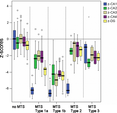
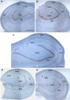
We propose a histopathological classification system for hippocampal cell loss in patients suffering from mesial temporal lobe epilepsies (MTLE). One hundred and seventy-eight surgically resected specimens were microscopically examined with respect to neuronal cell loss in hippocampal subfields CA1-CA4 and dentate gyrus. Five distinct patterns were recognized within a consecutive cohort of anatomically well-preserved surgical specimens. The first group comprised hippocampi with neuronal cell densities not significantly different from age matched autopsy controls [no mesial temporal sclerosis (no MTS); n = 34, 19%]. A classical pattern with severe cell loss in CA1 and moderate neuronal loss in all other subfields excluding CA2 was observed in 33 cases (19%), whereas the vast majority of cases showed extensive neuronal cell loss in all hippocampal subfields (n = 94, 53%). Due to considerable similarities of neuronal cell loss patterns and clinical histories, we designated these two groups as MTS type 1a and 1b, respectively. We further distinguished two atypical variants characterized either by severe neuronal loss restricted to sector CA1 (MTS type 2; n = 10, 6%) or to the hilar region (MTS type 3, n = 7, 4%). Correlation with clinical data pointed to an early age of initial precipitating injury (IPI < 3 years) as important predictor of hippocampal pathology, i.e. MTS type 1a and 1b. In MTS type 2, IPIs were documented at a later age (mean 6 years), whereas in MTS type 3 and normal appearing hippocampus (no MTS) the first event appeared beyond the age of 13 and 16 years, respectively. In addition, postsurgical outcome was significantly worse in atypical MTS, especially MTS type 3 with only 28% of patients having seizure relief after 1-year follow-up period, compared to successful seizure control in MTS types 1a and 1b (72 and 73%). Our classification system appears suitable for stratifying the clinically heterogeneous group of MTLE patients also with respect to postsurgical outcome studies.
Figures


References
-
- {'text': '', 'ref_index': 1, 'ids': [{'type': 'DOI', 'value': '10.1002/ana.410400314', 'is_inner': False, 'url': 'https://doi.org/10.1002/ana.410400314'}, {'type': 'PubMed', 'value': '8797534', 'is_inner': True, 'url': 'https://pubmed.ncbi.nlm.nih.gov/8797534/'}]}
- Arruda F, Cendes F, Andermann F, Dubeau F, Villemure JG, Jonesgotman M, Poulin N, Arnold DL, Olivier A (1996) Mesial atrophy and outcome after amygdalohippocampectomy or temporal lobe removal. Ann Neurol 40:446–450 - PubMed
-
- {'text': '', 'ref_index': 1, 'ids': [{'type': 'DOI', 'value': '10.1046/j.1528-1157.2001.43300.x', 'is_inner': False, 'url': 'https://doi.org/10.1046/j.1528-1157.2001.43300.x'}, {'type': 'PubMed', 'value': '11879344', 'is_inner': True, 'url': 'https://pubmed.ncbi.nlm.nih.gov/11879344/'}]}
- Bien CG, Kurthen M, Baron K, Lux S, Helmstaedter C, Schramm J, Elger CE (2001) Long-term seizure outcome and antiepileptic drug treatment in surgically treated temporal lobe epilepsy patients: a controlled study. Epilepsia 42:1416–1421 - PubMed
-
- {'text': '', 'ref_index': 1, 'ids': [{'type': 'DOI', 'value': '10.1007/s004010050952', 'is_inner': False, 'url': 'https://doi.org/10.1007/s004010050952'}, {'type': 'PubMed', 'value': '9930892', 'is_inner': True, 'url': 'https://pubmed.ncbi.nlm.nih.gov/9930892/'}]}
- Blumcke I, Beck H, Suter B, Hoffmann D, Födisch HJ, Wolf HK, Schramm J, Elger CE, Wiestler OD (1999) An increase of hippocampal calretinin-immunoreactive neurons correlates with early febrile seizures in temporal lobe epilepsy. Acta Neuropathol 97:31–39 - PubMed
-
- {'text': '', 'ref_index': 1, 'ids': [{'type': 'DOI', 'value': '10.1002/hipo.1045', 'is_inner': False, 'url': 'https://doi.org/10.1002/hipo.1045'}, {'type': 'PubMed', 'value': '11769312', 'is_inner': True, 'url': 'https://pubmed.ncbi.nlm.nih.gov/11769312/'}]}
- Blumcke I, Schewe JC, Normann S, Brustle O, Schramm J, Elger CE, Wiestler OD (2001) Increase of nestin-immunoreactive neural precursor cells in the dentate gyrus of pediatric patients with early-onset temporal lobe epilepsy. Hippocampus 11:311–321 - PubMed
-
- {'text': '', 'ref_index': 1, 'ids': [{'type': 'PMC', 'value': 'PMC8095862', 'is_inner': False, 'url': 'https://pmc.ncbi.nlm.nih.gov/articles/PMC8095862/'}, {'type': 'PubMed', 'value': '11958375', 'is_inner': True, 'url': 'https://pubmed.ncbi.nlm.nih.gov/11958375/'}]}
- Blumcke I, Thom M, Wiestler OD (2002) Ammon’s horn sclerosis: a maldevelopmental disorder associated with temporal lobe epilepsy. Brain Pathol 12:199–211 - PMC - PubMed
Publication types
MeSH terms
Substances
LinkOut - more resources
Full Text Sources
Other Literature Sources
Medical
Miscellaneous

