

Bisphenol A exposure in utero disrupts early oogenesis in the mouse
- PMID: 17222059
- PMCID: PMC1781485
- DOI: 10.1371/journal.pgen.0030005
Bisphenol A exposure in utero disrupts early oogenesis in the mouse
Abstract
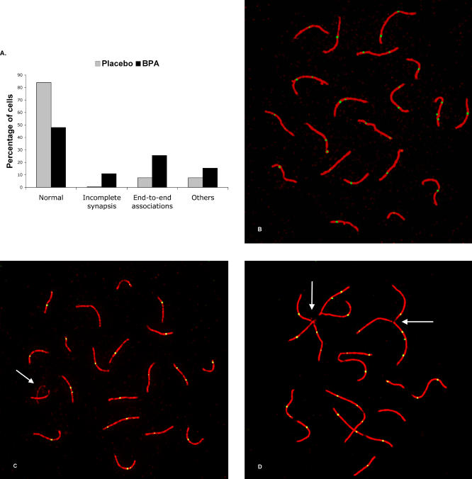
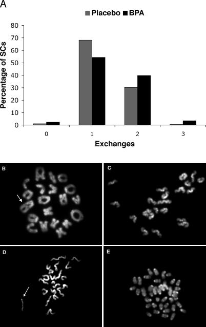
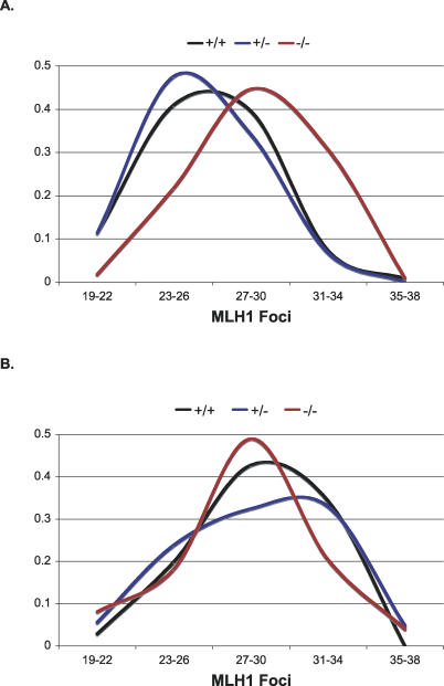
Estrogen plays an essential role in the growth and maturation of the mammalian oocyte, and recent studies suggest that it also influences follicle formation in the neonatal ovary. In the course of studies designed to assess the effect of the estrogenic chemical bisphenol A (BPA) on mammalian oogenesis, we uncovered an estrogenic effect at an even earlier stage of oocyte development--at the onset of meiosis in the fetal ovary. Pregnant mice were treated with low, environmentally relevant doses of BPA during mid-gestation to assess the effect of BPA on the developing ovary. Oocytes from exposed female fetuses displayed gross aberrations in meiotic prophase, including synaptic defects and increased levels of recombination. In the mature female, these aberrations were translated into an increase in aneuploid eggs and embryos. Surprisingly, we observed the same constellation of meiotic defects in fetal ovaries of mice homozygous for a targeted disruption of ERbeta, one of the two known estrogen receptors. This, coupled with the finding that BPA exposure elicited no additional effects in ERbeta null females, suggests that BPA exerts its effect on the early oocyte by interfering with the actions of ERbeta. Together, our results show that BPA can influence early meiotic events and, importantly, indicate that the oocyte itself may be directly responsive to estrogen during early oogenesis. This raises concern that brief exposures during fetal development to substances that mimic or antagonize the effects of estrogen may adversely influence oocyte development in the exposed female fetus.
Conflict of interest statement
Competing interests. The authors have declared that no competing interests exist.
Figures
Comment in
-
Scrambling eggs in plastic bottles.PLoS Genet. 2007 Jan;3(1):e6. doi: 10.1371/journal.pgen.0030006. PLoS Genet. 2007. PMID: 17222060 Free PMC article. No abstract available.
Similar articles
-
Human meiotic progression and recombination are affected by Bisphenol A exposure during in vitro human oocyte development.Hum Reprod. 2011 Oct;26(10):2807-18. doi: 10.1093/humrep/der249. Epub 2011 Jul 26. Hum Reprod. 2011. PMID: 21795248
-
Bisphenol a exposure causes meiotic aneuploidy in the female mouse.Curr Biol. 2003 Apr 1;13(7):546-53. doi: 10.1016/s0960-9822(03)00189-1. Curr Biol. 2003. PMID: 12676084
-
Bisphenol A alters early oogenesis and follicle formation in the fetal ovary of the rhesus monkey.Proc Natl Acad Sci U S A. 2012 Oct 23;109(43):17525-30. doi: 10.1073/pnas.1207854109. Epub 2012 Sep 24. Proc Natl Acad Sci U S A. 2012. PMID: 23012422 Free PMC article.
-
Bisphenol A Effects on Mammalian Oogenesis and Epigenetic Integrity of Oocytes: A Case Study Exploring Risks of Endocrine Disrupting Chemicals.Biomed Res Int. 2015;2015:698795. doi: 10.1155/2015/698795. Epub 2015 Aug 3. Biomed Res Int. 2015. PMID: 26339634 Free PMC article. Review.
-
Human female meiosis: what makes a good egg go bad?Trends Genet. 2008 Feb;24(2):86-93. doi: 10.1016/j.tig.2007.11.010. Epub 2008 Jan 14. Trends Genet. 2008. PMID: 18192063 Review.
Cited by
-
Altered cohesin gene dosage affects Mammalian meiotic chromosome structure and behavior.PLoS Genet. 2013;9(2):e1003241. doi: 10.1371/journal.pgen.1003241. Epub 2013 Feb 7. PLoS Genet. 2013. PMID: 23408896 Free PMC article.
-
The bisphenol S contamination level observed in human follicular fluid affects the development of porcine oocytes.Front Cell Dev Biol. 2023 Apr 6;11:1145182. doi: 10.3389/fcell.2023.1145182. eCollection 2023. Front Cell Dev Biol. 2023. PMID: 37091980 Free PMC article.
-
The bisphenol A experience: a primer for the analysis of environmental effects on mammalian reproduction.Biol Reprod. 2009 Nov;81(5):807-13. doi: 10.1095/biolreprod.109.077008. Epub 2009 May 20. Biol Reprod. 2009. PMID: 19458313 Free PMC article. Review.
-
Evaluating the Effects of BPA and TBBPA Exposure on Pregnancy Loss and Maternal-Fetal Immune Cells in Mice.Environ Health Perspect. 2022 Mar;130(3):37010. doi: 10.1289/EHP10640. Epub 2022 Mar 28. Environ Health Perspect. 2022. PMID: 35343813 Free PMC article.
-
Effects of Endocrine-Disrupting Chemicals on the Ovary.Biol Reprod. 2015 Jul;93(1):20. doi: 10.1095/biolreprod.115.130336. Epub 2015 Jun 10. Biol Reprod. 2015. PMID: 26063868 Free PMC article. Review.
References
-
- Herbst AL, Scully RE. Adenocarcinoma of the vagina in adolescence. Cancer. 1970;25:745–747. - PubMed
-
- Herbst AL. Behavior of estrogen-associated female genital tract cancer and its relation to neoplasia following intrauterine exposure to diethylstilbestrol (DES) Gynecol Oncol. 2000;76:147–156. - PubMed
-
- Newbold RR, Padilla-Banks E, Jefferson WN. Adverse effects of the model environmental estrogen diethylstilbestrol are transmitted to subsequent generations. Endocrinology. 2006;147:S11–S17. - PubMed
-
- Ikezuki Y, Tsutsumi O, Takai Y, Kamei Y, Taketani Y. Determination of bisphenol A concentrations in human biological fluids reveals significant early prenatal exposure. Hum Reprod. 2002;17:2839–2841. - PubMed
Publication types
MeSH terms
Substances
LinkOut - more resources
Full Text Sources
Other Literature Sources
Molecular Biology Databases
