Comparative Study
. 2007 Jan;274(1):275-86.
doi: 10.1111/j.1742-4658.2006.05585.x.

Crystal structures of a bacterial 6-phosphogluconate dehydrogenase reveal aspects of specificity, mechanism and mode of inhibition by analogues of high-energy reaction intermediates

Affiliations
Comparative Study

Crystal structures of a bacterial 6-phosphogluconate dehydrogenase reveal aspects of specificity, mechanism and mode of inhibition by analogues of high-energy reaction intermediates

Ramasubramanian Sundaramoorthy et al. FEBS J. 2007 Jan.

Abstract

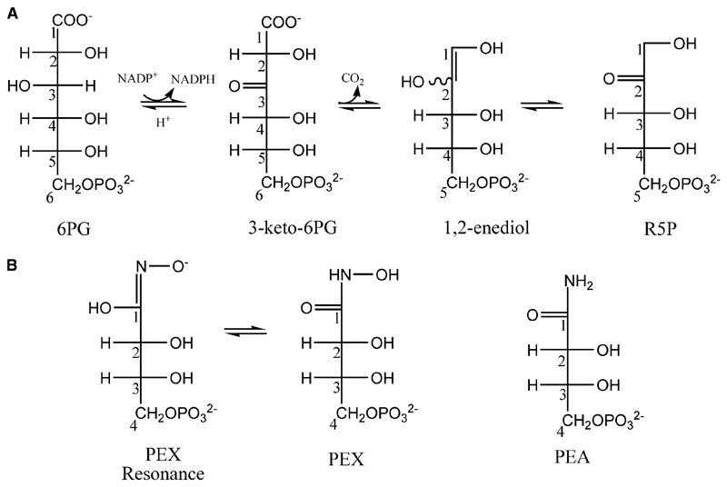
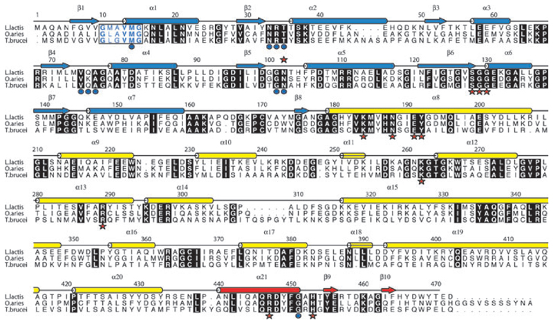
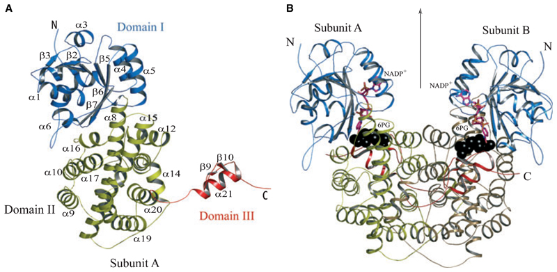
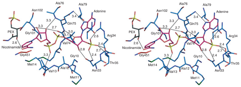
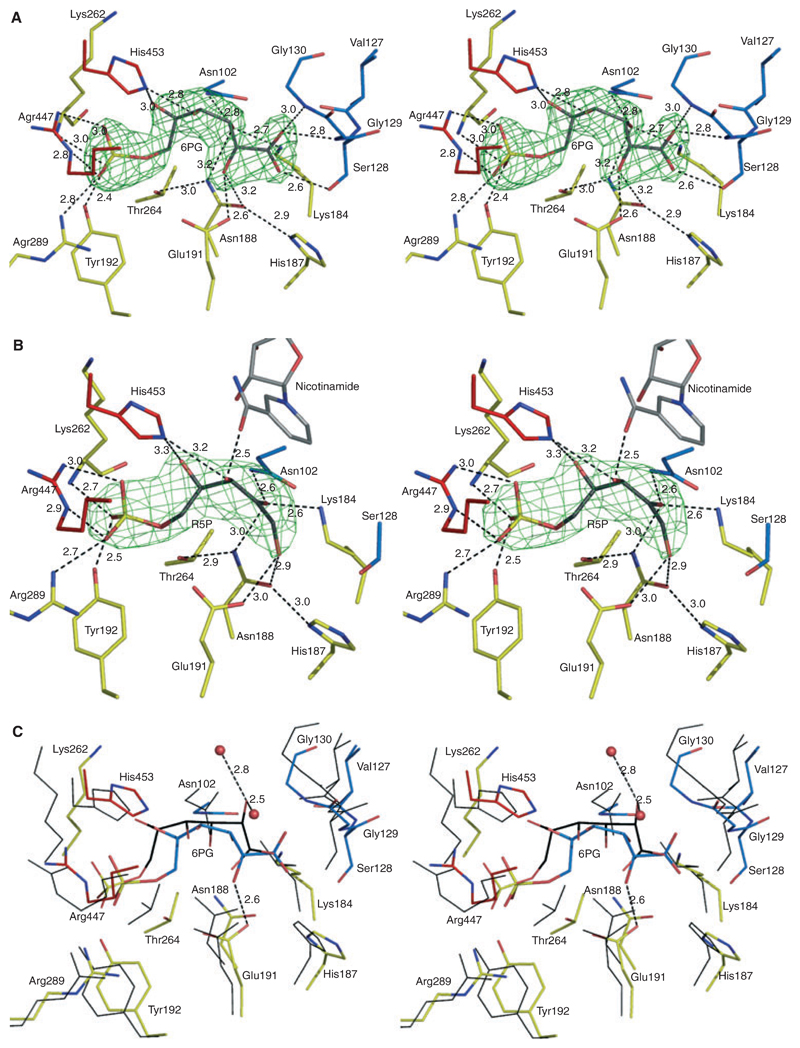
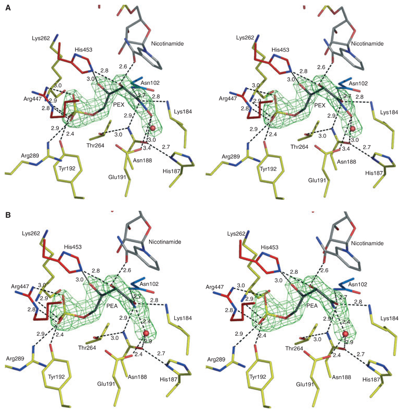
Crystal structures of recombinant Lactococcus lactis 6-phosphogluconate dehydrogenase (LlPDH) in complex with substrate, cofactor, product and inhibitors have been determined. LlPDH shares significant sequence identity with the enzymes from sheep liver and the protozoan parasite Trypanosoma brucei for which structures have been reported. Comparisons indicate that the key residues in the active site are highly conserved, as are the interactions with the cofactor and the product ribulose 5-phosphate. However, there are differences in the conformation of the substrate 6-phosphogluconate which may reflect distinct states relevant to catalysis. Analysis of the complex formed with the potent inhibitor 4-phospho-d-erythronohydroxamic acid, suggests that this molecule does indeed mimic the high-energy intermediate state that it was designed to. The analysis also identified, as a contaminant by-product of the inhibitor synthesis, 4-phospho-d-erythronamide, which binds in similar fashion. LlPDH can now serve as a model system for structure-based inhibitor design targeting the enzyme from Trypanosoma species.


Figures

Fig. 1
Fig. 1
(A) Catalytic reaction of PDH and two intermediate states. (B) Structures and numbering of the inhibitors PEX (two resonance forms) and PEA.
Fig. 2
Fig. 2
Amino acid sequence and secondary structure of LlPDH. Arrows depict β strands, cylinders depict α helices and these are labelled β1–β10 and α1–α21. The elements of secondary structure are coloured according to the domain in which they occur; blue for domain I, yellow for domain II and red for domain III. Aligned sequences of OaPDH and TbPDH are also shown. Most of the amino acids conserved in all three sequences are shown as white letters in black boxes. The exception is the NADP+ fingerprint region (residues 10–15 in LlPDH) where the letters are coloured blue and bold indicates conservation. Red stars identify active site residues that form direct hydrogen bonding interaction with ligands, blue dots identify those residues that interact with cofactor.
Fig. 3
Fig. 3
(A) Ribbon diagram of an LlPDH subunit. Elements of secondary structure are coloured according to domain as described in Fig. 2 and labelled. The N- and C-termini are marked. (B) The LlPDH dimer viewed perpendicular to the molecular twofold axis of symmetry, which is marked by an arrow. Black spheres depict the position of the substrate (6PG) at the catalytic centre, a stick model is shown for NADP+ and the cofactor is colored according to atom type; C is pink, N is blue, O is orange and P is yellow.
Fig. 4
Fig. 4
Stereoview of the cofactor-binding site in complex III. NADP+ is shown as in Fig. 3. LlPDH C atoms are blue, PEX C atoms are black and NADP+ C atoms are pink. All N positions are dark blue, O are red, P are yellow and S are green. Potential hydrogen bonding interactions are depicted as black dashed lines with interatomic distances given in Å.
Fig. 5
Fig. 5
Stereoviews showing interactions at the catalytic centre of LlPDH. (A) The omit difference density map (green mesh) for the substrate 6PG is shown. The map was calculated with coefficients |FoFc|, αcalc and contoured at 4σ. Fo and Fc represent observed and calculated structure-factor amplitudes, respectively, αcalc phases calculated on the basis of atomic coordinates of the model but excluding the substrate. 6PG atomic positions are coloured are follows: C, grey; O, red; P, yellow. The amino acid C atoms are coloured by domain assignment as in Fig. 2. Domain I is blue, domain II is yellow and domain III is red. O positions are red, N are blue. Black dashed lines represent potential hydrogen bonds with distances given in Å. (B) Binding of product, RU5P with the associated omit map. (B) is similar to (A), though note the presence of NADP+ with C atoms grey. (C) The superposition of LlPDH : 6PG and OaPDH : 6PG complexes based on Cα positions of residues shown. LlPDH : 6PG is shown as in (A) except that the C positions of 6PG are coloured cyan. Thin black lines represent OaPDH residues and the associated 6PG is shown as a stick model with C atoms in black.
Fig. 6
Fig. 6
Stereoviews depicting inhibition of LlPDH. (A) Omit difference density map (green mesh) in the active site calculated as described in Fig. 5 by ignoring the scattering contributions from the ligands and the water (red sphere) in estimating αcalc, the map is contoured at 4σ. The PEX model is shown with C atoms in black. (B) PEA model.

References

    1. Barrett MP. The pentose phosphate pathway and parasitic protozoa. Parasitol Today. 1997;13:11–16. - PubMed
    1. Hanau S, Rinaldi E, Dallocchio F, Gilbert IH, Dardonville C, Adams MJ, Gover S, Barrett MP. 6-Phosphogluconate dehydrogenase: a target for drugs in African trypanosomes. Curr Med Chem. 2004;11:2639–2650. - PubMed
    1. Gvozdev VA, Gerasimova TI, Kogan GL, Braslavskaya O. Role of the pentose phosphate pathway in metabolism of Drosophila melanogaster elucidated by mutations affecting glucose 6-phosphate and 6-phosphogluconate dehydrogenases. FEBS Lett. 1976;64:85–88. - PubMed
    1. Hughes MB, Lucchesi JC. Genetic rescue of a lethal ‘null’ activity allele of 6-phosphogluconate dehydrogenase in Drosophila melanogaster. Science. 1977;196:1114–1115. - PubMed
    1. Lobo Z, Maitra PK. Pentose phosphate pathway mutants of yeast. Mol Gen Genet. 1982;185:367–368. - PubMed

Publication types

MeSH terms