. 2007 Mar 2;145(1):56-65.
doi: 10.1016/j.neuroscience.2006.11.047. Epub 2007 Jan 10.

The actin binding domain of ACF7 binds directly to the tetratricopeptide repeat domains of rapsyn

Affiliations

The actin binding domain of ACF7 binds directly to the tetratricopeptide repeat domains of rapsyn

C Antolik et al. Neuroscience. .

Abstract

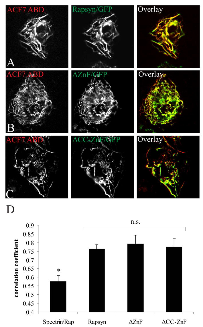
Formation of the neuromuscular junction requires the release of agrin from the presynaptic terminal of motor neurons. Clustering of acetylcholine receptors (AChRs) on the postsynaptic sarcolemma is initiated by agrin-dependent activation of the muscle-specific kinase. While the postsynaptic scaffolding protein rapsyn is vital for high density AChR aggregation, little is known about the mechanism through which AChRs are immobilized on the postsynaptic membrane. Ultrastructural and immunohistochemical studies of rat skeletal muscle have suggested that AChRs are anchored to a membrane-associated cytoskeleton that contains spectrin-like proteins and is thus similar to that of the human erythrocyte [Bloch RJ, Bezakova G, Ursitti JA, Zhou D, Pumplin DW (1997) A membrane skeleton that clusters nicotinic acetylcholine receptors in muscle. Soc Gen Physiol Ser 52:177-195]. We are studying a protein of the spectrin superfamily, ACF7 (also known as MACF), as a postsynaptic cytoskeletal component of the neuromuscular junction. ACF7 has multiple cytoskeleton-binding domains, including an N-terminal actin-binding domain that, we postulate, may interact with rapsyn, the scaffolding protein that binds directly to AChRs. To test this hypothesis, we co-expressed fragments of these molecules in cultured fibroblasts and assessed their co-distribution and interaction using confocal microscopy and co-immunoprecipitation. We demonstrate that the actin-binding domain of ACF7 specifically interacts with the tetratricopeptide repeat domains of rapsyn. Furthermore, we show using surface plasmon resonance and blot overlay that the actin-binding domain of ACF7 binds directly to rapsyn. These results suggest that, in mammalian skeletal muscle, AChRs are immobilized in the membrane through rapsyn-mediated anchoring to an ACF7-containing network that in turn is linked to the actin cytoskeleton.


Figures

Fig. 1
Fig. 1
The ABD of ACF7 specifically co-distributes with rapsyn in COS cells. Cells were co-transfected with plasmids encoding rapsyn/GFP (green) and DsRed-tagged constructs (red) encoding the ABDs of ACF7 (A), β-spectrin (B), dystrophin (C), utrophin (D), and filamin (E). Cells were also transfected with the ACF7 ABD construct alone and actin was labeled with phalloidin (green; F). As a control, rapsyn/GFP alone was expressed in COS cells (G). Cells were fixed 24 h later and fluorescent proteins and phalloidin were visualized with confocal microscopy. Rapsyn/GFP was recruited into actin filaments when co-expressed with the ACF7 ABD (A; areas of co-localization are shown in yellow in the overlay panel), but not the other ABDs (B–E).
Fig. 2
Fig. 2
Rapsyn’s TPR domains mediate co-distribution with the ACF7 ABD in COS cells. Cells co-expressing the ABD of ACF7 and full length rapsyn/GFP (A), rapsyn ΔZnF/GFP (B) or rapsyn ΔCC-ZnF/GFP (C) were fixed and imaged as in Fig. 1. The ABD of ACF7 (red) and each rapsyn construct (green) co-distributed significantly in actin filaments (yellow in the overlay panel). (D) Quantitative analysis showed that there was no difference in co-localization with the ABD of ACF7 and each of the rapsyn constructs, but that all three constructs showed significantly higher co-distribution than the ABD of spectrin with full length rapsyn (mean ± S.E.M., n= 20 from 2–3 separate experiments). n.s., not significantly different (p > 0.1, one-way ANOVA followed by Tukey’s HSD post hoc test); *, spectrin ABD is significantly different from ACF7 ABD co-distribution with rapsyn constructs (p < 0.01); Rap, full length rapsyn.
Fig. 3
Fig. 3
The ACF7 ABD interacts biochemically with the TPR domains of rapsyn. Lysates from cells co-transfected with plasmids encoding DsRed-tagged ACF7 ABD and either full length rapsyn/GFP, ΔZnF/GFP or ΔCC-ZnF/GFP were immunoprecipitated with anti-GFP antibodies. Following SDS-PAGE, precipitates were immunoblotted with anti-DsRed antibodies. The ACF7 ABD was specifically detected in immunoprecipitates from cells expressing rapsyn constructs, while the spectrin ABD was not detected in immunoprecipitates of cells co-expressing full length rapsyn/GFP. Immunoblots of lysates with anti-GFP and anti-DsRed showed that rapsyn and ABD constructs expressed at similar levels (inputs).
Fig. 4
Fig. 4
The ABD of ACF7 co-distributes with TPR domain pairs. COS cells were co-transfected with plasmids encoding DsRed-tagged ACF7 ABD and GFP constructs containing TPR domains 1–2 (A), 2–3 (B), 3–4 (C), 4–5 (D), 5–6 (E) and 6–7 (F). Cells were processed for fluorescence and imaged as in Fig. 1. The ACF7 ABD (red) and each rapsyn TPR pair (green) co-localized significantly (yellow in the overlay panel). (G) Quantitative analysis confirmed that there was no difference in the co-distribution of the ACF7 ABD with any of the TPR pairs, and that all of these combinations resulted in co-distributions that were significantly greater than with GFP alone (mean ± S.E.M., n= 20 from 2–3 separate experiments). n.s., not significantly different (p > 0.1, one-way ANOVA followed by Tukey’s HSD post hoc test); *, GFP control is significantly different from rapsyn constructs (p < 0.05).
Fig. 5
Fig. 5
The ABD of ACF7 binds directly to rapsyn. (A) Recombinant MBP and MBP-tagged ABD proteins were purified on amylose resins, separated by SDS-PAGE (1.4 μg and 2 μg per lane, respectively) and stained with Coomassie Blue. (B) Rapsyn/GFP and GFP alone were immunoprecipitated with anti-GFP antibodies from extracts of transfected COS cells, separated by SDS-PAGE and transferred to nitrocellulose. Blots were then incubated with recombinant MBP-tagged ACF7 ABD (left panel) or MBP alone (middle panel). After washing, bound recombinant proteins were detected with anti-MBP antibodies and chemiluminescence. MBP-tagged ACF7 ABD bound directly to rapsyn/GFP but not GFP alone; MBP did not bind to rapsyn/GFP. Immunoblotting confirmed the presence of equivalent amounts of GFP-tagged protein in each sample (right panel). (C) Rapsyn/GFP and GFP alone were purified as above and immobilized with anti-GFP antibodies on Biacore sensor chips. Recombinant MBP-tagged ACF7 ABD, at concentrations from 0.5 to 2 μM, was used as soluble binding partner. The y-axis shows binding in Resonance Units (RU) as a function of time after injection of recombinant ABD. Each curve represents binding at a specific concentration of recombinant ABD. Non-specific binding was subtracted from the binding of ABD to rapsyn.
Fig. 6
Fig. 6
The ABD of ACF7, but not the spectrin repeat region, is closely associated with rapsyn in situ. Rat myotubes in culture were sheared open and fixed (A–C, A′–C′), treated with 2 M NaCl and fixed (D–F, D′–F′), or treated with 2 M NaCl followed by 500 ng/ml chymotrypsin and fixed (G–I, G′–I′). Samples were labeled for immunofluorescence with monoclonal antibodies to rapsyn (B,B′, E,E′, H,H′) and affinity purified antibodies to the actin binding domain (A, D, G) or to the sixth spectrin repeat (A′, D′, G′) of ACF7. Both antibodies to ACF7 labeled rapsyn-rich structures in freshly isolated and fixed samples (A–C, A′–C′), and in samples treated briefly with 2 M NaCl (D–F, D′–F′). Epitopes in the actin binding domain of ACF7 (G–I), but not the spectrin repeat region (G′–I′) are retained after mild proteolysis with chymotryspin. Bar, 5 μm.
Fig. 7
Fig. 7
The ACF7 ABD, rapsyn, and MuSK760–820 co-distribute in COS cells. (A) Cells were triple-transfected with plasmids encoding DsRed-tagged ACF7 ABD, full length rapsyn/GFP and MuSK760–820/HA. After 24 h, cells were fixed and processed for immunocytochemistry with anti-HA antibodies, followed by Alexa-633-conjugated secondary antibodies. Images were obtained with the Kr/Ar (red and green) and He/Ne (blue) laser excitation. DsRed-tagged ACF7 ABD (red), full length rapsyn/GFP (green) and MuSK760–820/HA (blue) co-distributed in filaments (yellow arrows; areas of white in overlay panel). (B) Cells were transfected with plasmids encoding DsRed-tagged ACF7 ABD and MuSK760–820/HA. After 24 h, cells were fixed and processed for immunocytochemistry with anti-HA antibodies, followed by Alexa-488-conjugated secondary antibodies. The ACF7 ABD and MuSK760–820/HA did not co-distribute in the absence of rapsyn co-expression.

References

    1. Antolik C, Ursitti JA, Resneck WG, O’Neill AM, Lee PC, Pumplin DW, Bloch RJ. Abstract Viewer/Itinerary Planner. Washington, DC: Society for Neuroscience; 2006a. ACF7, a β-spectrin-like protein at clusters of acetylcholine receptors, associates with rapsyn. Program No. 30.2. 2006 Online.
    1. Antolik C, Catino DH, Resneck WG, Bloch RJ. The tetratricopeptide repeat domains of rapsyn bind directly to cytoplasmic sequences of the muscle-specific kinase. Neuroscience. 2006b;141:87–100. - PubMed
    1. Avnur Z, Geiger B. Substrate-attached membranes of cultured cells isolation and characterization of ventral cell membranes and the associated cytoskeleton. J Mol Biol. 1981;153:361–379. - PubMed
    1. Bartoli M, Ramarao MK, Cohen JB. Interactions of the rapsyn RING-H2 domain with dystroglycan. J Biol Chem. 2001;276:24911–24917. - PubMed
    1. Bezakova G, Bloch RJ. The zinc finger domain of the 43-kDa receptor-associated protein, rapsyn: role in acetylcholine receptor clustering. Mol Cell Neurosci. 1998;11:274–288. - PubMed

Publication types

MeSH terms