Review
. 2007:69:99-165.
doi: 10.1016/S0065-3527(06)69003-9.

The baculoviruses occlusion-derived virus: virion structure and function

Affiliations
Review

The baculoviruses occlusion-derived virus: virion structure and function

Jeffery Slack et al. Adv Virus Res. 2007.

Abstract

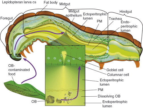
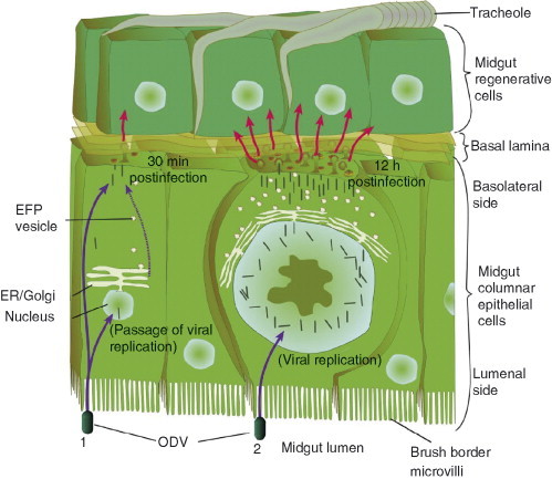
Baculoviruses play an important ecological role regulating the size of insect populations. For many years, baculoviruses have been applied as targeted biocontrol agents against forestry and agriculture pests. Baculovirus insecticides are effective against insect pests such as velvetbean caterpillar (Anticarsia gemmatalis), cotton bollworm (Helicoverpa zea), and gypsy moth (Lymantria dispar). Baculoviruses are transmitted to insects by the oral route mediated by the occlusion-derived virus (ODV). The ODV is also specialized to exploit the insect midgut that is one of the most extreme biological environments where the viruses are subject to caustic pH and digestive proteases. The molecular biology of the ODV reveals new frontiers in protein chemistry. Finally, ODVs establishes infection in insect gut tissues that are virtually nonsupportive to virus replication and which are continuously sloughed away. ODVs carry with them a battery of proteins that enable them to rapidly exploit and harness these unstable cells for virus replication.


Figures

Fig 1
Fig 1
Per os infection by baculoviruses. A cross‐sectional representation of the anatomy of an insect larva is depicted. A baculovirus OB enters by the per os route in contaminated food. OBs pass through the foregut and enter the midgut where they dissolve in the alkaline midgut lumen and release ODVs. The inset figure depicts the translocation of released ODVs past the peritrophic membrane (PM) to midgut columnar epithelial cells. The midgut region surrounded by the PM has been referred to as the endoperitrophic lumen and the region outside the PM has been referred to as the ectoperitrophic lumen.
Fig 2
Fig 2
Major occlusion‐derived virion forms. Three major OB phenotypes are illustrated in the background. The nucleopolyhedrovirus (NPV) OBs are larger than the Granulovirus (GV) OBs due to the fact that they contain multiple numbers of ODV virions. The OBs of GVs are capsule shaped and contain only single virions. The OBs of NPVs are multisided crystals or polyhedra. Some species of NPVs produce cuboidal OBs. The NPVs are further divided into the multiple nucleopolyhedroviruses (MNPVs) and single nucleopolyhedroviruses (SNPVs). The multiple (M) and single (S) designations are in reference to the number of nucleocapsids that are found in each virion. The ODVs of MNPVs, SNPVs, and GVs are depicted in the foreground. The ODVs are illustrated in dissected views and the GV ODV is illustrated as partially encapsulated.
Fig 3
Fig 3
The ODV phenotype. The illustration on the left side represents a dissected view of the structure of the ODV. The DNA genome is shown expanding from the nucleocapsid to emphasize the presence of one viral genome in each nucleocapsid. This illustration is done in the context of an MNPV ODV. In the background and expanded into the center are representations of the insect midgut epithelium. The ODV‐specific processes of attachment and viral envelope fusion with membranes of the brush border microvilli are shown. Expanded on the right hand side is resulting the release of ODV‐nucleocapsids which are translocated up the microvillus into the midgut cell.
Fig 4
Fig 4
The BV phenotype. The illustration on the left side represents a dissected view of the structure of the BV. The DNA genome is shown expanding from the nucleocapsid in order to emphasize its presence in the nucleocapsid. The major BV envelope fusion proteins (EFPs), GP64 and F protein are shown at the upper peplomer end of the virion. This illustration is in the context of a group I NPV baculovirus. The right hand side illustrates the processes of BV egress from an infected cell (lower right) and BV infection of a new cell (upper right). Nucleocapsids bud out of the infected cell membrane where viral EFPs have concentrated. In budding, the virion acquires EFPs and the cell membrane as its virion envelope. The BV diffuses across to a new cell where it is taken into the cell by receptor‐mediated endocytosis. The BV‐containing endosome fuses with an acidifying lysosome. This pH shift triggers EFP‐mediated envelope fusion with the endosomal membrane and release the BV nucleocapsid into the cytosol. The nucleocapsid then translocates to the nucleus.
Fig 5
Fig 5
Two modes of infection in the midgut. Most baculoviruses use the midgut only to produce the first generation of BV progeny that bud out from the basal lateral side of the midgut cell to infect other tissues. ODVs that enter midgut cells may (1) bypass virus replication in the midgut cell or (2) initiate virus replication. When MNPVs bypass replication, most nucleocapsids translocate to the basolateral side of the midgut cell. One or a few nucleocapsids enter the nucleus, unpackaged, and expresses envelope fusion protein (efp) genes. EFP proteins are translocated via the endoplasmic reticulum (ER) and Golgi to the basal lateral side of the cell where they meet up with nucleocapsids which then bud out. This is the passage effect. When MNPVs replicate in the midgut cell significantly greater numbers of BV are produced. However, this mode of infection requires 8–12 h vs 30 min when the virus bypasses viral replication.
Fig 6
Fig 6
Baculovirus infection cycle. Several phases of virus replication are illustrated beginning with the rounding of newly infected cells and finishing with the lytic release of OBs. Indicated times are relative to the infection cycle of AcMNPV. The purpose of the figure is to illustrate the progression of phases from BV production to ODV production. Nucleocapsids are initially translocated to the cell membrane for BV production and later become retained in the nuclear ring zone for ODV production. INM is in reference to the inner nuclear membrane (INM) which provides the ODV envelope.
Fig 7
Fig 7
Proteins of the ODV nucleocapsid. Some of the proteins found associated with the ODV nucleocapsid are illustrated. The figure is in the context of AcMNPV. The “*” indicates that the proteins for which the specific location on the nucleocapsids has not been confirmed. We have also placed nucleocapsid‐associated proteins which have DNA‐binding activities or potential transcriptional activation activities on the viral genome. The intent of the figure is to reveal the nucleocapsid as a complex structure of multiple protein types instead of simply protein sheath surrounding Basic Protein and the viral genome.
Fig 8
Fig 8
Proteins of the ODV envelope and tegument. The illustration represents a dissected view of the structure of the ODV with emphasis on the envelope. The figure is in the context of AcMNPV. The major ODV‐associated proteins with predicted transmembrane domains are shown in the ODV envelope. The “*” indicates that there is no data to suggest the orientation of these proteins in the ODV envelope. FP25K, which is capsid associated, also appears on the ODV envelope due to its apparent interaction with the transmembrane domains of ODV‐E66 and ODV‐E25. Tegument proteins are also illustrated in the main virion on the right side.
Fig 9
Fig 9
A simple model for baculovirus virion envelopment and envelope protein translocation. The BV and ODV forms in distinctly different locations in the cell. BV nucleocapsid must translocate through nuclear pores out of the nucleus, across the cytosol, and to the cell membrane. ODV nucleocapsids are retained within the nuclear ring zone. The formation of enveloped virions in the nucleus is unusual for viruses. However, there may be a common thread between the ODV and the BV morphogenesis. Cell surface proteins such as BV integral envelope fusion proteins (EFPs) are synthesized in the ER such that they are exposed in the ER lumen. EFPs are translocated to the cell surface in ER vesicles, which after sorting in the Golgi complex, fuse with the host cell membrane. This permits EFPs to be presented on the outside of BVs as they bud out of the cell. ODV virions may acquire their envelopes by “budding” into INM vesicles. The double nuclear membrane is continuous with the ER. In this model, ODV envelope proteins such as P74 and F protein would be displayed on the luminal side of the ER and nuclear double membrane. Envelope proteins such as E66 that may interact with nucleocapsids would be displayed on the nucleoplasmic side of INM vesicles.

Similar articles

Cited by

References

    1. Abbas M., Boucias D.G. Interaction between nuclear polyhedrosis virus‐infected Anticarsia gemmatalis (Lepidoptera: Noctuidae) larvae and predator Podisus maculiventris (Say) (Hemiptera: Pentatomidae) Environ. Entomol. 1984;13(2):599–602.
    1. Aboussekhra A., Wood R.D. Detection of nucleotide excision repair incisions in human fibroblasts by immunostaining for PCNA. Exp. Cell Res. 1995;221(2):326–332. - PubMed
    1. Ackermann H.W., Smirnoff W.A. A morphological investigation of 23 baculoviruses. J. Invertebr. Pathol. 1983;41(3):269–280.
    1. Adams J.R., Bonami J.R. Atlas of invertebrate viruses. In: Adams J.R., Bonami J.R., editors. “Atlas of InvertebrateViruses”. CRC Press; Boca Raton: 1991.
    1. Adams J.R., McClintock J.T. Baculoviridae, nuclear polyhedrosis viruses Part 1. Nuclear polyhedrosis viruses of insects. In: Adams J.R., Bonami J.R., editors. “Atlas of Invertebrate Viruses”. CRC Press; Boca Raton: 1991. pp. 87–180. Chapter 6.

Publication types

Substances

LinkOut - more resources