

Review
doi: 10.1016/j.ccr.2006.12.008.
Tracking normalization of brain tumor vasculature by magnetic imaging and proangiogenic biomarkers
Affiliations
- PMID: 17222788
- PMCID: PMC2952447
- DOI: 10.1016/j.ccr.2006.12.008
Item in Clipboard
Review
Tracking normalization of brain tumor vasculature by magnetic imaging and proangiogenic biomarkers
Cancer Cell.
2007 Jan.
Abstract
Clinical assessment of the response to antiangiogenic therapy has been cumbersome. A study in this issue of Cancer Cell demonstrates that a combination of magnetic resonance imaging (MRI) for quantification of normalized vessels with measurements of circulating levels of proangiogenic factors, including FGF2, SDF1, and viable circulating endothelial cells, provides an effective means to evaluate the response of recurrent glioblastoma to a prototypical pan-VEGF receptor tyrosine kinase inhibitor, AZD2171.
Figures
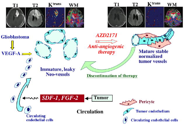
ZD2171 targets immature vessels, while favoring the emergence of normalized pericyte coated blood vessels. Recurrence of the tumor is associated with generation of dilated, large, and leaky blood vessels as well as plasma elevation of proangiogenic factors, including FGF2, SDF1, and CECs. Normalization of the blood vessels is associated with a decrease in post-contrast T1-weighted MRI signal (T1), T2-weighted (flair, T2), permeability (Ktrans), and recovery of white matter integrity as detected by white matter tractography (WM). The MRI photographs were adapted from Batchelor et al. (2007).
Comment on
-
AZD2171, a pan-VEGF receptor tyrosine kinase inhibitor, normalizes tumor vasculature and alleviates edema in glioblastoma patients.Cancer Cell. 2007 Jan;11(1):83-95. doi: 10.1016/j.ccr.2006.11.021. Cancer Cell. 2007. PMID: 17222792 Free PMC article. Clinical Trial.
References
-
- Cha S. Magn Reson Imaging Clin N Am. 2003;11:403–413. - PubMed
-
- Dennie J, Mandeville JB, Boxerman JL, Packard SD, Rosen BR, Weisskoff RM. Magn Reson Med. 1998;40:793–799. - PubMed
-
- Hegi ME, Diserens AC, Gorlia T, Hamou MF, de Tribolet N, Weller M, Kros JM, Hainfellner JA, Mason W, Mariani L, et al. N Engl J Med. 2005;352:997–1003. - PubMed
-
- Jain RK. Science. 2005;307:58–62. - PubMed
Publication types
MeSH terms
Substances
Grants and funding
LinkOut - more resources
Full Text Sources
Other Literature Sources
Medical
