. 2007 Mar 9;366(5):1510-22.
doi: 10.1016/j.jmb.2006.12.044. Epub 2006 Dec 21.

Relationships between the sequence of alpha-synuclein and its membrane affinity, fibrillization propensity, and yeast toxicity

Affiliations

Relationships between the sequence of alpha-synuclein and its membrane affinity, fibrillization propensity, and yeast toxicity

Michael J Volles et al. J Mol Biol. .

Abstract

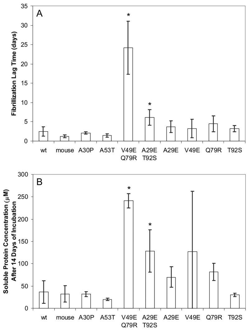
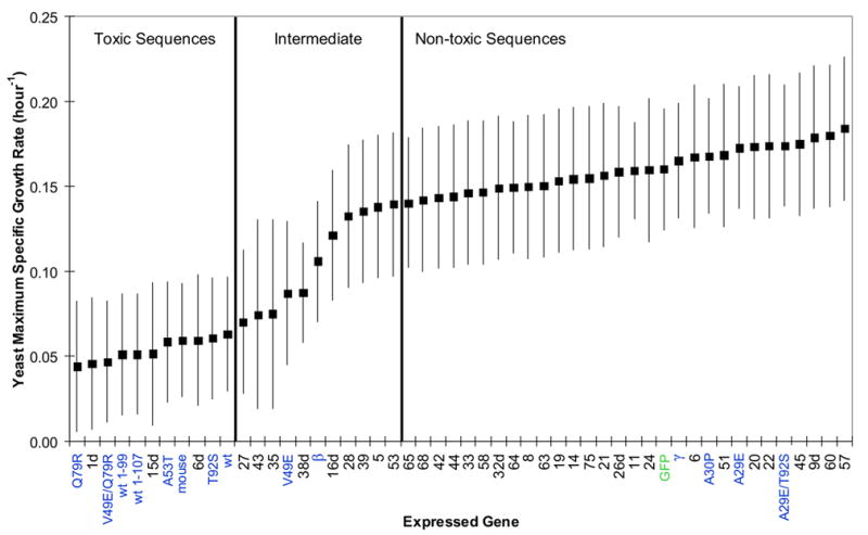
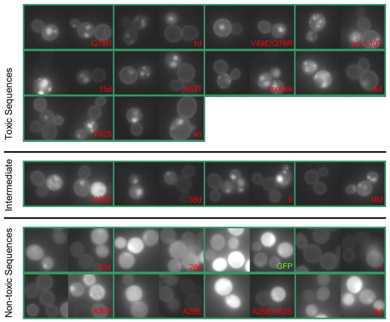
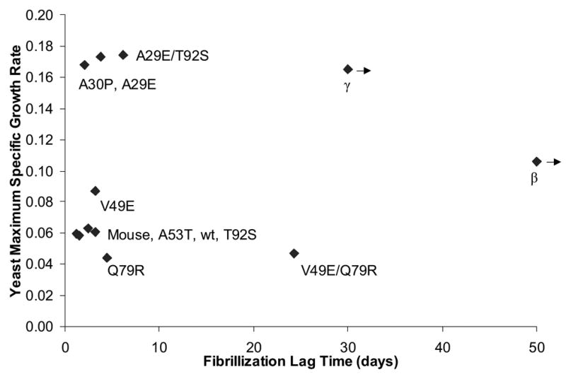
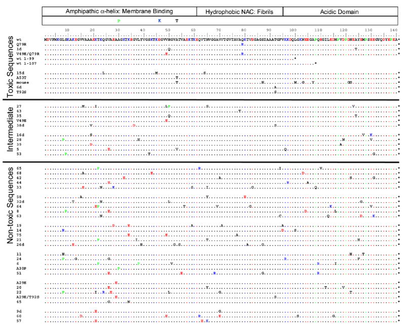
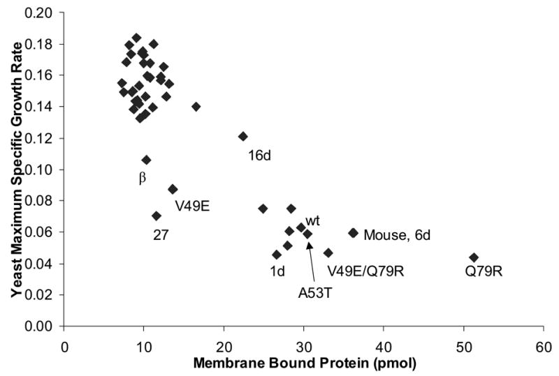
To investigate the alpha-synuclein protein and its role in Parkinson's disease, we screened a library of random point mutants both in vitro and in yeast to find variants in an unbiased way that could help us understand the sequence-phenotype relationship. We developed a rapid purification method that allowed us to screen 59 synuclein mutants in vitro and discovered two double-point mutants that fibrillized slowly relative to wild-type, A30P, and A53T alpha-synucleins. The yeast toxicity of all of these proteins was measured, and we found no correlation with fibrillization rate, suggesting that fibrillization is not necessary for synuclein-induced yeast toxicity. We found that beta-synuclein was of intermediate toxicity to yeast, and gamma-synuclein was non-toxic. Co-expression of Parkinson's disease-related genes DJ-1, parkin, Pink1, UCH-L1, or synphilin, with synuclein, did not affect synuclein toxicity. A second screen, of several thousand library clones in yeast, identified 25 non-toxic alpha-synuclein sequence variants. Most of these contained a mutation to either proline or glutamic acid that caused a defect in membrane binding. We hypothesize that yeast toxicity is caused by synuclein binding directly to membranes at levels sufficient to non-specifically disrupt homeostasis.


Figures

Figure 1
Figure 1
Fibrillization of control α-synuclein sequences, two double point mutant sequence variants, and their single point mutant derivatives. Each bar represents the mean of eight (except single point mutants, where n = 4) trials of independently expressed, purified, and incubated proteins, and the error bars are individual 95% confidence intervals. Single point mutants were created from the double point mutants by subcloning with the aid of enzyme BseRI. (A) Lag time is defined by the crossing of a thioflavin T fluorescence threshold as described in the methods. In 2 of 8 V49E/Q79R incubations, a positive thioflavin T signal had not occurred within the duration of the experiment. In these cases, the day of the final measurement was used as the lag time (32 days). Therefore the value shown for this variant may be somewhat underestimated. (B) Soluble protein concentrations measured after 14 days of incubation. Every non-V49E/Q79R sample had fibrillized within 8 days. Using a pre-planned t-test on the data of (A) or (B), we were able to reject the null hypothesis of no difference in lag time between the wild type protein and both V49E/Q79R and A29E/T92S with a p-value < 0.01 (indicated by an asterisk). A Games-Howell (variance is non-constant; reference51) multiple comparison procedure of (A) did not detect a significant difference between the four derived single point mutants and the wt protein or A29E/T92S. However, comparison of V49E/Q79R with any of the other proteins resulted in rejection of the hypothesis of no difference. Applying the Games-Howell test to the data in (B) gave similar results but was somewhat more liberal in rejecting the null hypothesis.
Figure 2
Figure 2
Estimated maximum specific growth rates (proportional to the inverse of doubling time) of the yeast strain W303-1a expressing various synuclein genes or GFP (labeled in green) from 2-micron plasmid p426GAL1. All genes isolated by yeast library screening are labeled numerically; those originally isolated from the yeast screen with co-expressed wt α-synuclein have numbers with the suffix “d”. All other synuclein sequences (those not isolated by yeast screening) have descriptive labels, shaded blue. The synuclein genes were not GFP tagged and all of the synuclein cDNAs were contained in the plasmid within the same non-coding nucleotide sequence context (except wt 1-107, whose C-terminal nucleotides after the stop codon were deleted). The genes are listed on the x-axis in order of decreasing estimated yeast toxicity. Each gene was studied in independent trials (n was typically about 6), independent meaning that a new trial was begun from a different colony picked from the same transformation. An additive 2-way ANOVA model (synuclein genes and blocks) was used for analysis of the maximum specific growth rates obtained by fitting the Gompertz equation. The square points are estimated population marginal means. In 2 of 306 samples sparse data led to a fit near 1.0, which was unreasonably high, and these outliers were set to a more reasonable upper limit of 0.3 to avoid disturbing the statistical analysis. Error bars represent simultaneous Tukey-Kramer comparison intervals: non-overlap between the bars of any two genes indicates that the hypothesis of no difference between the two was rejected at the p<0.05 level. Two vertical bars divide the sequences into three groups: “toxic”, “intermediate”, and “non-toxic”. Every sequence in the non-toxic group is significantly less toxic to yeast than every sequence in the toxic group. Sequences in the intermediate group have comparison intervals that overlap some members of both the toxic and non-toxic sets.
Figure 3
Figure 3
Microscopy of GFP tagged synucleins. Two horizontal bars divide the synuclein variants into three toxicity groups, as defined in Fig. 2. Photographs were taken after 48 hours of growth in synthetic galactose medium lacking uracil, and all were taken with identical excitation intensity and exposure time (100× plan fluor objective; a Gaussian filter was applied to the images; the levels and gamma setting were adjusted identically for all images). Each green box contains two different representative photographic fields and the labels correspond with those used elsewhere in the manuscript. The expression plasmid was p426GAL1 with GFP (the linkage between the last amino acid of synuclein and GFP is described in the methods section, and was identical for α, β, and γ synucleins); synuclein cDNAs were inserted into the plasmid by in vitro ligation.
Figure 4
Figure 4
Scatter plot of estimated maximum specific growth rate (data from Fig. 2) versus in vitro synuclein fibrillization lag time (data from Fig. 1), for a number of synuclein variants (labeled). Left-pointing arrows are shown for β and γ-synuclein to indicate that their true (unknown) lag times are actually greater than the value shown; in the case of β-synuclein, the true value is much greater.
Figure 5
Figure 5
α-Synuclein sequences studied by quantitative yeast growth are shown. The division of the α-synuclein sequence into domains is shown at the top. Just below this, the PD-linked mutations are shown. The wild type α-synuclein sequence is the first in the list, and other sequences are listed in order of decreasing estimated yeast toxicity from top to bottom; sequences are divided into three statistical groups as in Figs. 2 and 3. Negatively/positively charged amino-acids are shown in red/blue, and proline is shaded green. As discussed in the methods, only amino acids 8–130 are highly mutatable because of mutation correction at the termini by PCR primers. Stop codons are indicated by an asterisk.
Figure 6
Figure 6
Scatter plot of estimated maximum specific growth rate (data from Fig. 2) versus membrane binding level (data from Supplementary Fig. 5). Membrane binding level is the amount of synuclein that co-sedimented with vesicles during a centrifugation assay (see methods). One point is shown for each of the 47 variants listed in Supplementary Fig. 5 (some are labeled).

References

    1. Cookson MR. The biochemistry of Parkinson’s disease. Annu Rev Biochem. 2005;74:29–52. - PubMed
    1. Conway KA, Harper JD, Lansbury PT., Jr Accelerated in vitro fibril formation by a mutant alpha-synuclein linked to early-onset Parkinson disease. Nat Med. 1998;4:1318–20. - PubMed
    1. Spillantini MG, Schmidt ML, Lee VM, Trojanowski JQ, Jakes R, Goedert M. Alpha-synuclein in Lewy bodies. Nature. 1997;388:839–40. - PubMed
    1. Bussell R, Jr, Ramlall TF, Eliezer D. Helix periodicity, topology, and dynamics of membrane-associated alpha-synuclein. Protein Sci. 2005;14:862–72. - PMC - PubMed
    1. Davidson WS, Jonas A, Clayton DF, George JM. Stabilization of alpha-synuclein secondary structure upon binding to synthetic membranes. J Biol Chem. 1998;273:9443–9. - PubMed

Publication types

MeSH terms