. 2007 May 10;361(2):316-24.
doi: 10.1016/j.virol.2006.11.031. Epub 2007 Jan 16.

Herpes simplex virus type 1 tegument proteins VP1/2 and UL37 are associated with intranuclear capsids

Affiliations

Herpes simplex virus type 1 tegument proteins VP1/2 and UL37 are associated with intranuclear capsids

Michelle A Bucks et al. Virology. .

Abstract

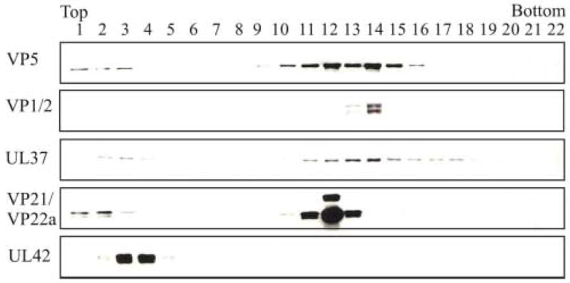
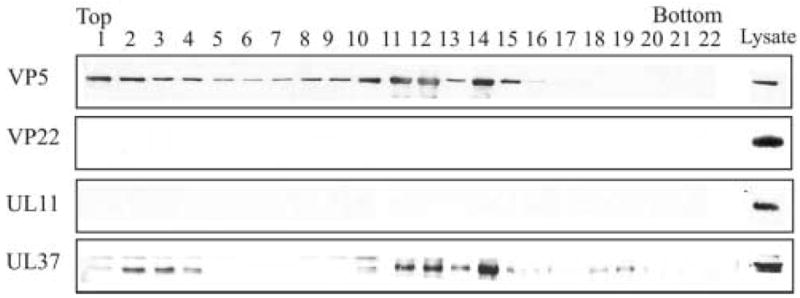
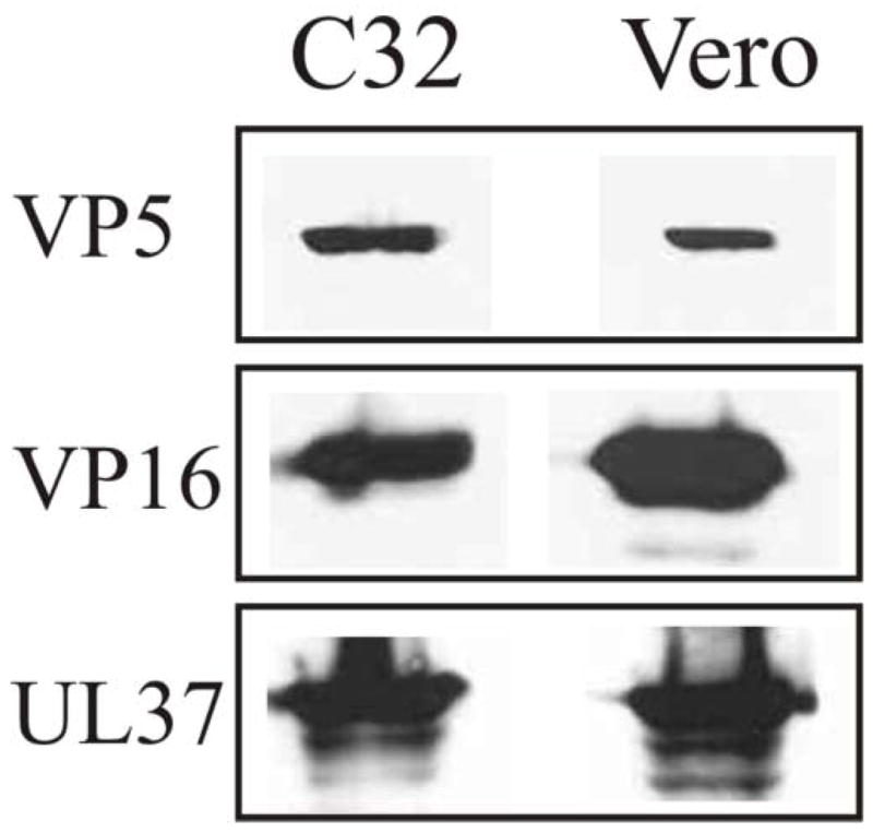
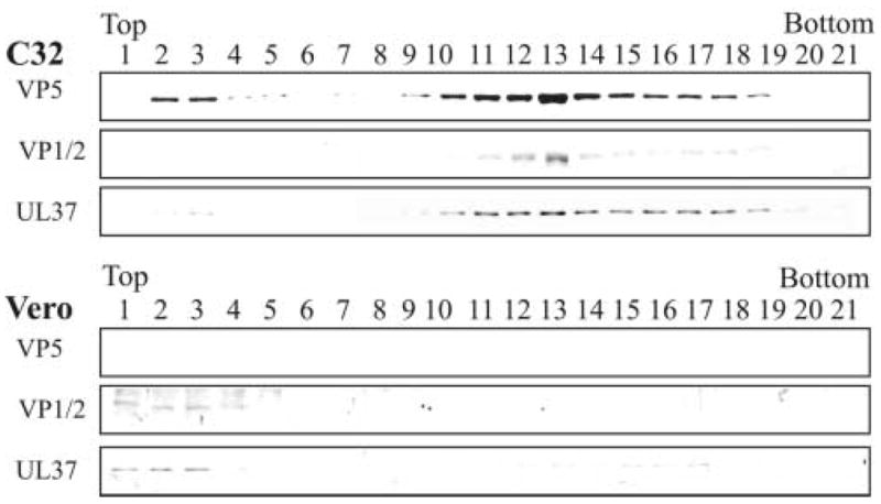
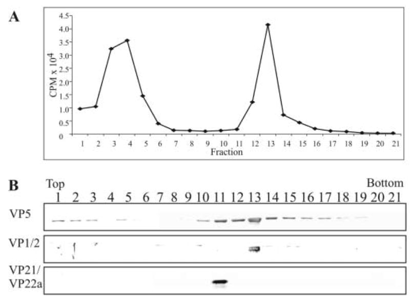
The assembly of the tegument of herpes simplex virus type 1 (HSV-1) is a complex process that involves a number of events at various sites within virus-infected cells. Our studies focused on determining whether tegument proteins, VP1/2 and UL37, are added to capsids located within the nucleus. Capsids were isolated from the nuclear fraction of HSV-1-infected cells and purified by rate-zonal centrifugation to separate B capsids (containing the scaffold proteins and no viral DNA) and C capsids (containing DNA and no scaffold proteins). Western blot analyses of these capsids indicated that VP1/2 associated primarily with C capsids and UL37 associated with B and C capsids. The results demonstrate that at least two of the tegument proteins of HSV-1 are associated with capsids isolated from the nuclear fraction, and these capsid-tegument protein interactions may represent initial events of the tegumentation process.


Figures

FIG. 1
FIG. 1
Analysis of the purity of the nuclear fraction obtained from HSV-1-infected cells. Approximately 2 x 10⁶ Vero cells were infected with HSV-1 at a MOI of 10 pfu/cell. At 15 h after infection, the cells were harvested and separated into the cytoplasmic and nuclear fractions as described in the Methods. The proteins of the nuclear and cytoplasmic fractions were resolved by SDS-PAGE followed by Western blot analysis simultaneously using antibodies to the nuclear marker lamin B1 (70 kDa) and the endoplasmic reticulum marker calnexin (90 kDa) to assay the same nitrocellulose blot.
FIG. 2
FIG. 2
Analysis of the purity of the nuclear capsid preparation. (A) Approximately 1.5 x 10⁸ HSV-1 KOS infected Vero cells were harvested at 15 h pi, resuspended in 1% NP40 lysis buffer and then mixed with the cytoplasmic fraction from approximately 1.3 x 10⁸ Vero cells infected with HSV-1 K26GFP virus. This mixture was incubated on ice for 30 minutes and the nuclear fraction was then purified as described in the Methods. Capsids from the nuclear fraction were purified and separated on a 20–50% linear sucrose gradient following rate-zonal centrifugation. 0.5 ml fractions were collected from the bottom of the tube. The proteins within each fraction were precipitated with 10% TCA, separated by SDS-PAGE followed by Western blot analysis. The blot was probed sequentially with antibodies to GFP and the major capsid protein VP5. K26GFP lysate (far right) shown in all panels served as a positive control for the Western blots. (B) Western blot analysis of capsids isolated from the cytoplasmic fraction from approximately 2.6 x 10⁷ K26GFP infected Vero cells purified and analyzed as described above. K26GFP lysate is shown in the far right lanes as a control.
FIG. 3
FIG. 3
Rate-zonal centrifugation of HSV-1 capsids isolated from the nuclear fraction and analyzed for the presence of tegument proteins. Capsids were obtained from the nuclear fraction of HSV-1-infected Vero cells harvested at 15 h after infection and separated on a 20–50% linear sucrose gradient following rate-zonal centrifugation. 0.5 ml fractions were collected from the bottom of the tube. The proteins within each fraction were precipitated with 10% TCA, separated by SDS-PAGE followed by Western blot analysis using primary antibodies to specific tegument proteins followed by the addition of the secondary anti-rabbit or anti-mouse antibody conjugated to horseradish peroxidase. Western blot analysis was done using antibodies to capsid proteins VP5 (the major capsid polypeptide) and VP21/VP22a (scaffold proteins), tegument proteins VP1/2 and UL37, and the DNA binding protein UL42.
FIG. 4
FIG. 4
Rate-zonal centrifugation of HSV-1 capsids isolated from the nuclear fraction and analyzed for the presence of tegument proteins VP22 and UL11. At 15 h after infection the capsids from the nuclear fraction were obtained, separated by rate-zonal centrifugation as described in the legend to Fig. 2. The same blot was probed sequentially using antibodies specific for the major capsid protein VP5 and tegument proteins VP22, UL11, and UL37 (positive control).
FIG. 5
FIG. 5
SDS-PAGE analysis of the cell lysates of Vero (VP23 non-complementing) and C32 (VP23 complementing) cells infected with the HSV-1 VP23-negative mutant virus, K23Z. The two cell lines were infected with the K23Z virus at a MOI of 10 pfu per cell and harvested at 15 h after infection. The whole cell lysates were resolved by SDS-PAGE followed by Western blot analysis. The same blot was probed sequentially with antibodies to VP5, VP16, and UL37.
FIG. 6
FIG. 6
Rate-zonal centrifugation of capsids isolated from cells infected with the VP23-negative mutant (K23Z) and analysis for the presence of tegument proteins VP1/2 and UL37. Vero (non-complementing cell line) and C32 (complementing cell line expressing VP23) cells were infected with the VP23-negative mutant virus, K23Z, at a MOI of 10 pfu/cell. At 15 h after infection the capsids from the nuclear fraction were obtained, separated by rate-zonal centrifugation, analyzed by SDS-PAGE followed by Western blot analysis as described in the legend to Fig. 2. The same blot was probed sequentially for the presence of VP5 and tegument proteins VP1/2 and UL37.
FIG. 7
FIG. 7
Rate-zonal centrifugation of 3H-thymidine-labeled capsids and analysis for the presence of the tegument protein VP1/2. Capsids were obtained from the nuclear fraction of HSV-1-infected Vero cells that were labeled with 10 μCi per ml of 3H-thymidine from 3 to 15 h after infection. The capsids were separated by rate-zonal centrifugation as described in the legend to Fig. 2. Prior to TCA precipitation of the proteins within each of the fractions, 50 μl was removed and 3H-thymidine cpm in each fraction were determined by liquid scintillation analysis (panel A). Following TCA precipitation, the same fractions were resolved by SDS-PAGE followed by Western blot analysis (panel B). The same blot was sequentially probed with antibodies to the tegument protein VP1/2 and capsid proteins VP5 and VP21/VP22a (scaffold proteins) as described in the legend to Fig. 2.

Similar articles

Cited by

References

    1. Albright AG, Jenkins FJ. The herpes simplex virus UL37 protein is phosphorylated in infected cells. J Virol. 1993;67 (8):4842–4847. - PMC - PubMed
    1. Batterson W, Furlong D, Roizman B. Molecular genetics of herpes simplex virus. VIII further characterization of a temperature-sensitive mutant defective in release of viral DNA and in other stages of the viral reproductive cycle. J Virol. 1983;45 (1):397–407. - PMC - PubMed
    1. Beard PM, Duffy C, Baines JD. Quantification of the DNA cleavage and packaging proteins U(L)15 and U(L)28 in A and B capsids of herpes simplex virus type 1. J Virol. 2004;78 (3):1367–1374. - PMC - PubMed
    1. Bowzard JB, Visalli RJ, Wilson CB, Loomis JS, Callahan EM, Courtney RJ, Wills JW. Membrane targeting properties of a herpesvirus tegument protein-retrovirus Gag chimera. J Virol. 2000;74 (18):8692–8699. - PMC - PubMed
    1. Chang YE, Van Sant C, Krug PW, Sears AE, Roizman B. The null mutant of the U(L)31 gene of herpes simplex virus 1: construction and phenotype in infected cells. J Virol. 1997;71 (11):8307–8315. - PMC - PubMed

Publication types

Substances