

Chronic wasting disease
- PMID: 17223321
- PMCID: PMC2680674
- DOI: 10.1016/j.bbadis.2006.10.010
Chronic wasting disease
Abstract
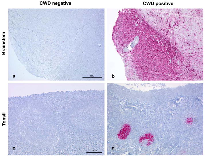
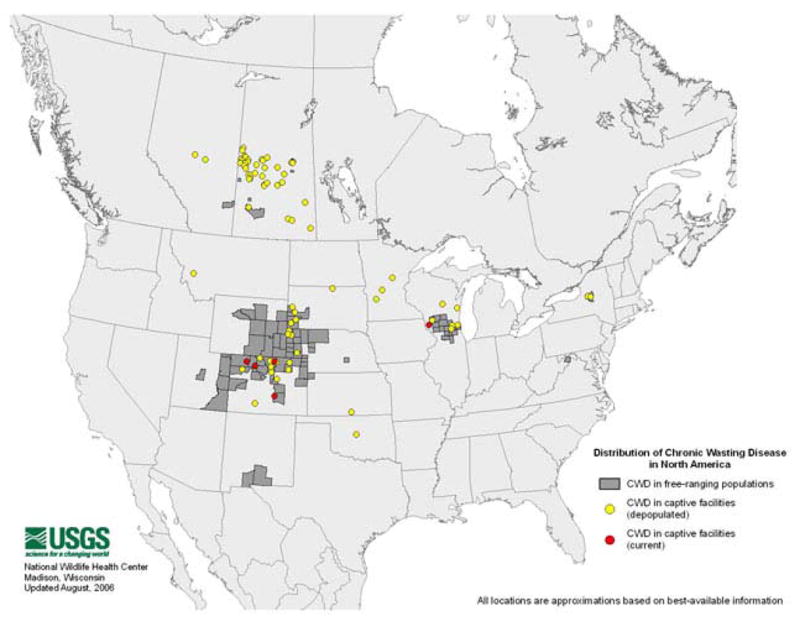
Until recently, chronic wasting disease of cervids, the only prion disease affecting wildlife, was believed to be geographically concentrated to Colorado and Wyoming within the United States. However, increased surveillance has unveiled several additional pockets of CWD-infected deer and elk in 12 additional states and 2 Canadian provinces. Deer and elk with CWD have extensive aggregates of PrP(Sc) not only in the central nervous system, but also in peripheral lymphoid tissues, skeletal muscle, and other organs, perhaps influencing prion shedding. Indeed, CWD is transmitted efficiently among animals by horizontal routes, although the mechanism of spread is unknown. Genetic polymorphisms in the Prnp gene may affect CWD susceptibility, particularly at codon 225 (S/F) in deer and codon 132 (M/L) in elk. Since CWD infects free-ranging animals and is efficiently spread, disease management will be a challenge.
Figures
References
-
- Williams ES, Miller MW. Chronic wasting disease in deer and elk in North America. Rev Sci Tech. 2002;21:305–16. - PubMed
-
- Williams ES. Chronic wasting disease. Vet Pathol. 2005;42:530–49. - PubMed
-
- Williams ES, Young S. Chronic wasting disease of captive mule deer: a spongiform encephalopathy. J Wildl Dis. 1980;16:89–98. - PubMed
-
- Spraker TR, Zink RR, Cummings BA, Wild MA, Miller MW, O’Rourke KI. Comparison of histological lesions and immunohistochemical staining of proteinase-resistant prion protein in a naturally occurring spongiform encephalopathy of free-ranging mule deer (Odocoileus hemionus) with those of chronic wasting disease of captive mule deer. Vet Pathol. 2002;39:110–9. - PubMed
Publication types
MeSH terms
Substances
Grants and funding
LinkOut - more resources
Full Text Sources
Research Materials
